
The anatomy of colorectal cancer staging
Staging colorectal cancer is the process of mapping exactly how far the disease has spread, and that map determines everything about your treatment.
Think of it like a structural inspection of a building. A colonoscopy tells your care team what is there; staging imaging tells them how far it has reached. Those are two very different questions requiring very different tools. A colonoscopy identifies and biopsies a suspicious lesion, but it can’t reveal whether cancer cells have traveled to nearby lymph nodes or distant organs.
The TNM framework – Tumor, Node, Metastasis – is the standard language oncologists use to answer those questions. Each letter captures a distinct dimension of the disease. The size and depth of the primary tumor (T), whether regional lymph nodes are involved (N), and whether metastasis to distant sites has occurred (M). Together, these three variables produce a final stage from 0 to 4, a shorthand your care team uses to communicate prognosis and plan next steps.
That stage number carries real weight. A patient with stage 3 colorectal cancer faces a very different sequence of treatment than someone at stage 2, often requiring chemotherapy before or after surgery rather than surgery alone. Staging isn’t a formality. It’s the clinical foundation that shapes every treatment decision that follows. And the imaging scans your care team orders are the instruments that make that precision possible, starting with the wide-angle view a CT scan provides.
CT scans: the wide-angle lens for body mapping
After a colorectal cancer screening flags a concern or if you have symptoms, your care team may recommend imaging tests such as CT scans to provide detailed information about the size and location of the cancer and whether it has spread.
The scan works by rotating an X-ray source around your body, capturing hundreds of thin cross-sectional “slices” that a computer then assembles into a detailed 3D map. That map covers the chest, abdomen, and pelvis simultaneously, which matters enormously when staging colorectal cancer. Knowing whether disease has stayed local or traveled elsewhere shapes every treatment decision that follows.
Detecting metastasis is the CT scan’s primary job in this context. The liver and lungs are the two most common sites where colorectal cancer spreads first. A chest CT scan is often used to check for lung metastases in patients with colorectal cancer, while separate imaging is used to evaluate the liver. This is especially relevant when evaluating stage 4 colorectal cancer outcomes, since distant metastasis is precisely what defines that stage and drives prognosis.
Local invasion is the other critical variable the scan measures. If a tumor has pushed through the colon wall into surrounding fatty tissue – a shift that moves the disease from Stage 2 toward more advanced classification – the CT can often reveal that breach. However, it’s worth noting a practical caveat. CT provides excellent broad coverage, but its soft-tissue contrast has limits, particularly in the pelvis. That’s exactly why rectal tumors often require a more precise tool, one built specifically to interrogate the fine anatomical detail that CT can miss.
MRI: The high-definition zoom for rectal precision
MRI is often used to help evaluate rectal tumors and may provide detailed images of the tumor and surrounding tissue. Your care team can discuss whether MRI is appropriate for your situation.
Evidence-based guidance powered by NCCN Guidelines®
Personalized treatment plans shaped by the latest oncology standards—tailored to your diagnosis.
Get started
View your personalized treatment plan in the Outcomes4Me app
Use your diagnosis to unlock personalized NCCN Guidelines®-aligned recommendations.
Continue in app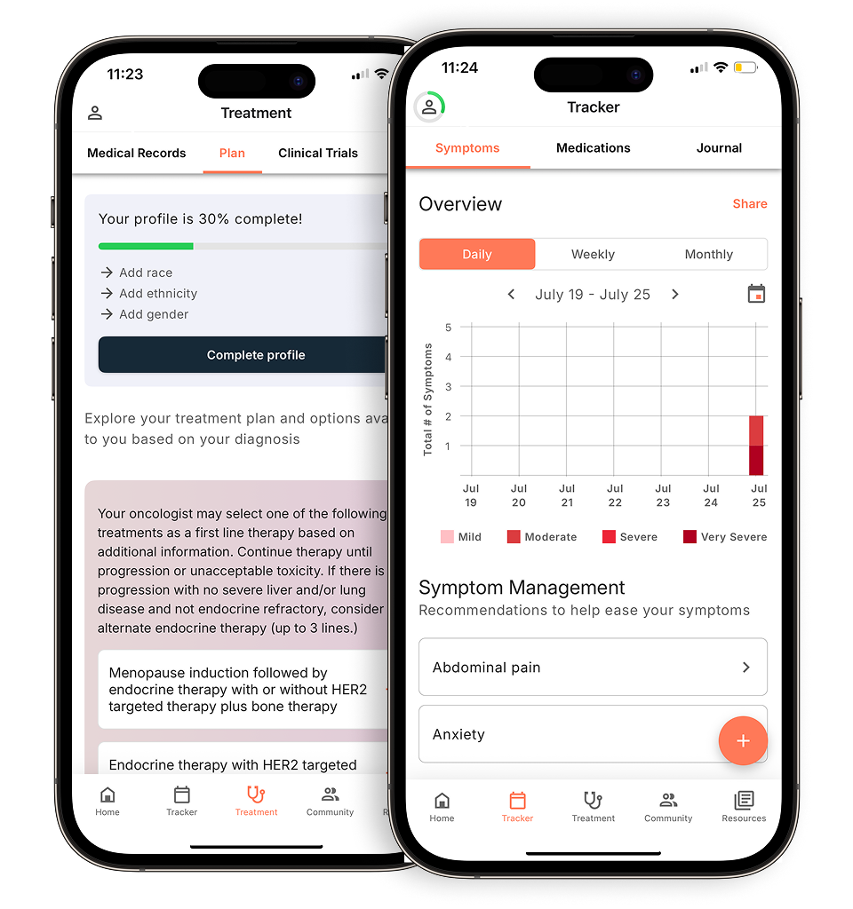
While CT scanning gives your care team a broad anatomical overview, MRI has superior soft-tissue contrast, making it the preferred modality for rectal cancer staging. That superior soft-tissue contrast lets radiologists trace exactly where a tumor ends and healthy tissue begins, a distinction that’s critical for surgical planning.
Circumferential Resection Margin (CRM) is one of the most important metrics MRI measures. CRM is a prognostic factor in rectal cancer that can be detected by high-resolution MRI and is linked to increased five-year survival and decreased risk of recurrence. A margin that’s too narrow signals a higher risk of cancer remaining after surgery.
MRI also evaluates the mesorectum, a layer of fat surrounding the rectum that doctors may examine during staging. Cancer cells that spread into the mesorectal tissue can travel along lymphatic channels. Mesorectal involvement is a key factor in both staging and personalizing care plans.
Each of these data points, margin status and mesorectal involvement, feeds directly into staging decisions. And as you’ll see in the next section, placing MRI alongside CT and PET scans in a direct comparison reveals exactly where each modality excels.
Comparison table: imaging modalities side-by-side
Choosing the right scan depends on what your care team needs to see, and CT, MRI, and PET each answer a different clinical question at staging.
The distinction between anatomical imaging (CT and MRI) and functional imaging (PET) is especially important when staging progresses toward stage 4 colon cancer, where distant spread must be confirmed precisely.
| Scan | Primary use | Detail level | Best anatomical target |
|---|---|---|---|
| CT | Full-body spread mapping | Moderate soft tissue | Liver metastases, lung nodules |
| MRI | Local tumor depth and margins | High soft tissue | Rectal wall layers, lymph nodes |
| PET | Metabolic activity detection | Functional, not structural | Distant or occult metastases |
Liver vs. lymph nodes: MRI is superior to CT for detecting liver metastases in colorectal cancer, although CT colonography can help with overall tumor staging. MRI edges ahead for lymph node assessment near the rectum, where soft-tissue contrast resolves small nodal detail that CT can miss.
The table above sets the foundation, but there are also specialized scans go even further, targeting specific anatomical scenarios that standard imaging can’t fully address.
What labs will be abnormal with colon cancer?
Blood tests don’t stage colon cancer on their own, but they surface biological signals that guide your care team toward the right imaging and treatment decisions.
Understanding what colorectal cancer does to your bloodwork helps explain why labs are ordered alongside scans at every stage. Three markers tend to shift most meaningfully.
Carcinoembryonic antigen (CEA) is the primary blood marker used in colorectal cancer management. Elevated CEA levels before surgery can correlate with more advanced disease. Rising CEA levels after treatment often signal recurrence before it’s visible on imaging. CEA is not a standalone diagnostic tool, but it adds an important layer to staging response monitoring.
Cancer care guidance for every step of your journey
Get treatment options, clinical trials, and support tailored to your diagnosis--all in one place.
Get started
See treatment options, manage symptoms, and stay informed—all in the app
View treatment options and trials personalized to your diagnosis—plus track progress in real time.
Continue in appComplete blood count (CBC) findings, particularly low hemoglobin, frequently point to chronic, slow bleeding from a tumor in the colon wall. Many patients first ask what is colorectal cancer only after a routine CBC flags unexplained anemia. That single result can be the earliest indirect sign of disease.
Liver function tests (LFTs) round out the picture when metastatic spread is a concern. Elevated enzymes like AST, ALT, or alkaline phosphatase may suggest the liver is under stress. Elevated liver enzymes are a red flag that prompts dedicated liver imaging to confirm or rule out metastases.
Taken together, these markers help your care team build a fuller clinical picture before and after scans are interpreted. And since CT remains the backbone of staging, the next question worth exploring is how well it actually detects earlier-stage disease.
Can a CT scan detect colon cancer in early stages?
CT scans are powerful staging tools, but they have limitations in detecting certain tumors.
Early-detection limits. At Stage 0 or Stage 1, cancer cells may be confined to a small polyp or the innermost mucosal layer. Standard CT scans have difficulty accurately staging colorectal cancer because they have poor soft-tissue contrast resolution, which is why CT colonography is used to improve tumor staging.
Why colonoscopy leads. CT scans may miss some colorectal tumors, particularly smaller or flat tumors, which is why colonoscopy remains the definitive diagnostic tool. A colonoscopy lets your care team directly visualize the mucosal lining, identify and biopsy suspicious polyps, and in many cases remove them on the spot. CT steps in after a colonoscopy confirms cancer, forming the staging baseline that maps how far the disease has traveled.
Virtual colonoscopy as an alternative. For patients who can’t tolerate sedation, take blood thinners, or have anatomical barriers to a standard scope, CT Colonography offers a middle ground. CT Colonography generates detailed 3D images of the colon interior without requiring a scope. CT Colonography is less invasive than standard colonoscopy, but if a polyp is found, a follow-up standard colonoscopy is still needed to remove it. Understanding these trade-offs is exactly the kind of logistical detail your care team will walk you through, which brings us to what the full staging workup actually looks like in practice.
The logistics of the workup: what to expect
The staging workup typically includes a biopsy to confirm the diagnosis and imaging tests to determine how far the cancer has spread.
The staging timeline typically unfolds in phases. A colonoscopy biopsy comes first, establishing that cancer is present. Imaging tests such as CT scans, MRI scans, or PET scans may be done to look at suspicious areas, learn how far cancer has spread, or help determine if treatment is working. In many cancer centers, staging information is reviewed by a multidisciplinary team of oncologists, radiologists, and surgeons who work together to interpret findings and help guide treatment planning.
Contrast agents play a bigger role in that imaging phase than most patients anticipate. Oral barium coats the inner lining of the colon to improve wall visibility. IV iodine contrast, injected during the scan, may help highlight blood vessels and distinguish tumor tissue from surrounding structures.
Clinical vs. pathological staging is where results can shift. The stage assigned before surgery, based entirely on imaging, is the clinical stage. After surgery, pathologists examine the removed tissue directly, and that analysis sometimes reveals more extensive disease. Stage migration can occur when additional information from surgery or pathology findings leads to a revised cancer stage classification after the initial assessment.
Even with sophisticated imaging protocols, scans don’t always tell the complete story, and that’s worth understanding before you interpret your own results.
Limitations and considerations in imaging
Imaging is a powerful diagnostic tool, but no scan delivers a perfect picture, and understanding where it falls short helps you ask better questions of your care team.
False positives and negatives are a real limitation. Inflammatory conditions like diverticulitis or post-surgical changes may appear similar to tumor activity on a CT or MRI, which is why your care team may recommend additional imaging or tests to clarify findings. Standard imaging has limitations in detecting lymph node involvement, particularly when relying on size-based measurements alone.
Patient-specific factors can also determine which scan is even possible. Significantly reduced kidney function may rule out contrast-enhanced CT, since the dye used is processed renally. Metal implants, pacemakers, or certain surgical hardware can make MRI unsafe or produce image artifacts that reduce diagnostic accuracy.
The “wait-and-see” period is another layer many patients don’t anticipate. When neoadjuvant therapy, such as radiation or chemotherapy for stage 4 colon cancer and locally advanced rectal disease, is used before surgery, scans are typically repeated afterward to assess tumor response. Restaging after neoadjuvant therapy is critical because it determines whether the tumor has shrunk enough for a less invasive resection or whether the treatment approach needs to shift. Taken together, these limitations reinforce why imaging is always interpreted in context, a point that ties directly into the broader takeaways for navigating your staging scans.
Key takeaways: navigating your staging scans
Staging is not a single test, it’s a coordinated process where CT scans map distant spread, MRI defines local surgical detail, and blood work like CEA adds critical context that imaging alone can’t provide.
Understanding how these pieces fit together helps you have more informed conversations with your care team. Abnormal labs such as low hemoglobin or an elevated CEA don’t deliver a verdict on their own, they’re signals that guide which imaging follows and how urgently. Staging findings help determine your colon cancer treatment plan, which may include surgery as the main treatment, chemotherapy or immunotherapy before or after surgery, or a combination of these approaches depending on how far the cancer has spread.
Stage has a direct bearing on the colorectal cancer cure rate. Early-stage disease caught before distant spread carries significantly better outcomes than advanced disease. Getting the correct diagnosis at the start of treatment matters, and it involves multiple steps including imaging tests and genetic testing to understand where your cancer started, its type, and whether it has spread.
Disclaimer: The information provided in this article is for informational purposes only and is not intended to be a substitute for professional medical advice, diagnosis, or treatment. Outcomes4Me is not acting as your caregiver, and any suggestions or guidance offered should not replace the advice of your healthcare provider or qualified medical professional. Always seek the guidance of your physician or other qualified health provider with any questions you may have regarding a medical condition.
Personalized support for real care decisions
Understand your diagnosis, explore clinical trials, and track symptoms--all in one place.
Get started
Compare treatments, prepare for appointments, and track side effects—all in the app
Built for your diagnosis, Outcomes4Me gives you the tools to make confident, informed decisions—right when you need them.
Continue in app