
On National Mammography Day, observed every year on the third Friday of October, we’re spotlighting one of the many tools used for breast cancer detection: mammograms. National Mammography Day serves as a reminder to schedule your mammogram or encourage a loved one to do so.
Alongside mammograms, other important screening methods, such as breast ultrasounds and MRIs, are also used for imaging. Understanding the different imaging techniques for breast cancer can help you and your team identify abnormalities, evaluate risks, and shape treatment strategies. This guide aims to simplify some of these tools, offering clarity and support.
Mammograms: 2D and 3D (Tomosynthesis)
A 2D mammogram is the traditional X-ray imaging technique of the breast and has been the gold standard for breast cancer screening for decades. It works by taking two flat images of each breast. While highly effective as an imaging technique in detecting breast cancer early, it has limitations, especially in women with dense breast tissue, where the overlapping layers can obscure or mimic cancerous growths.
Evidence-based guidance powered by NCCN Guidelines®
Personalized treatment plans shaped by the latest oncology standards—tailored to your diagnosis.
Get started
View your personalized treatment plan in the Outcomes4Me app
Use your diagnosis to unlock personalized NCCN Guidelines®-aligned recommendations.
Continue in app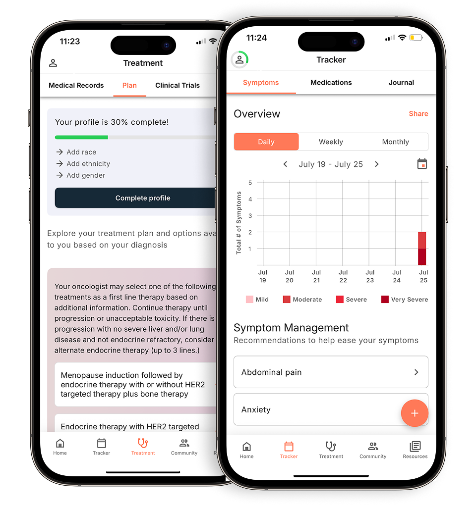
Tomosynthesis, or 3D mammography, is a more recent innovation that takes multiple X-ray pictures of each breast from different angles, creating a layered, three-dimensional image. This method allows doctors to see through layers of tissue and examine areas of concern more clearly, reducing the need for follow-up testing and false positives. It’s particularly beneficial for individuals with dense breast tissue.
Screening mammograms are routine for those without symptoms as part of regular health check-ups to catch cancer early. Diagnostic mammograms are performed if you or your doctor find breast changes or if a screening mammogram detects an irregularity. They are more detailed and focus on a specific area of the breast.
As of September 10th, 2024, the Food and Drug Administration (FDA) mandates that all mammogram reports sent to patients must include information about breast density. This is a crucial development because dense breast tissue can make it harder to detect cancer on a mammogram, as both dense tissue and tumors appear white. Additionally, having dense breasts can increase the risk of developing breast cancer.
Including this information empowers patients and healthcare providers to make more informed decisions. For those with dense breasts, other screening methods such as breast ultrasounds or MRIs might be recommended to ensure more comprehensive monitoring and detection.
Breast ultrasound
Breast ultrasound uses sound waves to produce images of the structures inside the breast. Unlike mammography, which uses X-rays, ultrasound provides a different type of image that can distinguish between fluid-filled cysts and solid masses.
Ultrasounds are often used alongside mammograms, especially in women with dense breast tissue, to evaluate abnormal findings from a mammogram or physical exam.
They are also often used if a biopsy is needed, as they help the doctor perform needle biopsies by providing real-time images.
Breast MRI
A breast MRI is an imaging technique that uses magnetic fields to create detailed pictures of the inside of the breasts to screen for cancer. It’s a more sensitive imaging test that can pick up more abnormalities than mammograms or ultrasounds but also has a higher rate of false positives.
For those with a strong family history of breast cancer or a genetic predisposition, MRIs can be used in addition to mammograms for comprehensive screening.
They are also often used to further evaluate abnormalities detected by a mammogram or ultrasound and to assess the extent of cancer after diagnosis.
Understanding the differences between these screening and diagnostic tools and their specific applications can help you navigate your breast health care more confidently. It’s essential to have open conversations with your healthcare provider about which tests are right for you, based on your individual risk factors and health history.
——————————————————————
Facing a breast cancer diagnosis can feel overwhelming, especially when trying to understand the maze of breast imaging options and what they mean for your treatment. Outcomes4Me is your partner in demystifying breast cancer care, providing you with the latest, evidence-based information right at your fingertips.
Download the Outcomes4Me app today and take a proactive step towards managing your breast health with knowledge and support.
Personalized support for real care decisions
Understand your diagnosis, explore clinical trials, and track symptoms--all in one place.
Get started
Compare treatments, prepare for appointments, and track side effects—all in the app
Built for your diagnosis, Outcomes4Me gives you the tools to make confident, informed decisions—right when you need them.
Continue in app