The following questions and responses have been lightly edited for grammatical purposes.
1) What’s the difference between 3D mammograms (breast tomosynthesis) and traditional 2D mammograms?
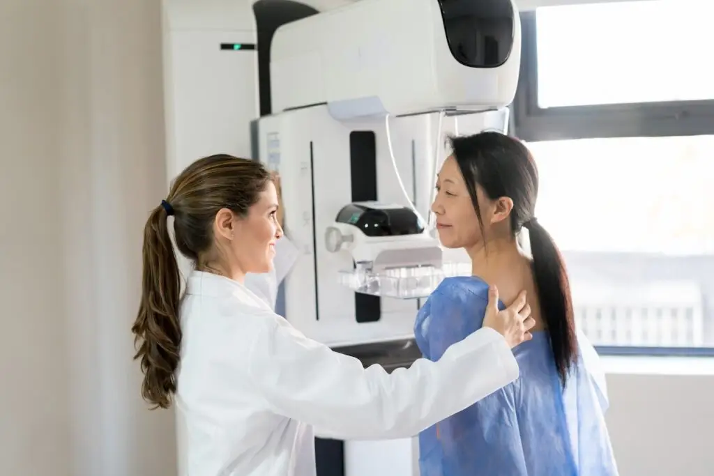
Dr. Amy Kelly: So 3D, otherwise known as digital breast tomosynthesis, is what we’re typically using these days to image the breast tissue. Most facilities are using digital breast tomosynthesis. The difference between a 3D mammogram and a 2D mammogram is that the 3D mammogram takes little slices through the breast tissue and allows us to really get a good look at the tissue. It reduces the masking effect of dense breast tissue because we’re able to take little slices through the breast tissue and it eliminates a lot, not all, of the problems with dense breast tissue and how it can mask or hide cancer.
2D imaging is just a two-dimensional picture. You’re not actually able to look through the dense tissue. We’ve been using 3D mammography for some time now, probably since 2012. What we know from all the research that we’ve done is it finds more cancers and there’s an improved cancer detection rate. One of the best things is that 3D mammography has a lower false-positive rate than 2D mammography. We’re actually calling fewer patients back,[and] we’re recommending fewer unnecessary benign biopsies with 3D versus 2D, so it’s great.
Evidence-based guidance powered by NCCN Guidelines®
Personalized treatment plans shaped by the latest oncology standards—tailored to your diagnosis.
Get started
View your personalized treatment plan in the Outcomes4Me app
Use your diagnosis to unlock personalized NCCN Guidelines®-aligned recommendations.
Continue in app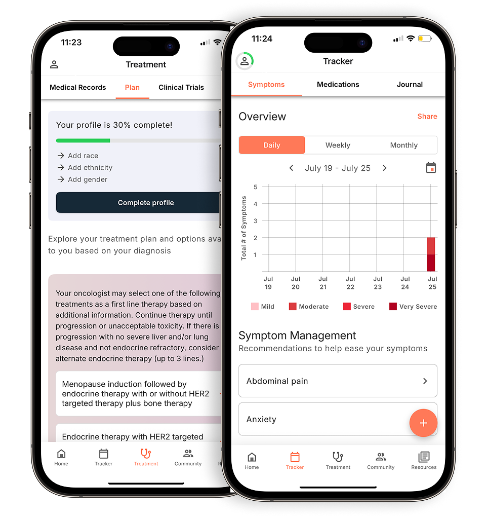
We really think that all patients should have it. It’s better for women with dense breasts. The benefit ratio is better for women with dense breast tissue versus those who have really fatty breast tissue, but even in a woman who does not have dense breast tissue, we are finding that it’s better than 2D mammography. We do recommend it for all women, especially women with dense tissue or who are at elevated risk.
2) What is contrast-enhanced mammography?
Dr. Amy Kelly: With contrast-enhanced mammography, what we do is inject intravenous (IV) iodinated contrast and then we take mammogram pictures both before and after the contrast is injected. This gives us valuable information about whether anything is enhancing. Ultimately, this technology is amazing. The sensitivity, specificity, positive predictive value, and negative predictive value are all superior to traditional mammography.
The sensitivity of detecting breast cancer with contrast-enhanced mammography is extraordinarily high, about 98%. This is similar to what we see with breast MRI, which is currently the most sensitive imaging tool we have for detecting breast cancer. So if cancer is there, MRI, and now contrast-enhanced mammography, are going to detect it.
One of the great things about contrast-enhanced mammography is that its false-positive rate is lower than that of MRI. While MRI is excellent, it can sometimes detect benign findings that lead to unnecessary biopsies. Contrast-enhanced mammography, on the other hand, finds fewer of these benign things, so it has a lower false-positive rate. It’s really checking all the boxes for us and is an excellent tool.
It’s FDA-approved to be used in the diagnostic setting for patients with signs and symptoms of breast cancer or those already being evaluated at our institution. We’re also using it in a research trial for women at very high risk for breast cancer, such as those with a personal history.
I want to mention that as physicians, breast imagers, and caregivers, we want the best imaging modalities to be as widely available and affordable as possible. The great thing about contrast-enhanced mammography is that it’s more accessible than breast MRI. Not every facility has an MRI magnet, and not every patient can undergo an MRI, so this is a fantastic alternative for those reasons.
3) How do I know if my imaging center is using the most up-to-date technology, like 3D mammography?
Dr. Amy Kelly: Most imaging facilities are using 3D. I do recommend, if you’re in more of a rural community, calling your facility and asking them if they do 3D mammography. Also ask them if they have supplemental screening options such as ultrasound, MRI, or even contrast-enhanced mammography if you have dense breast tissue.
If you’re calling them and asking about their availability of technology, I do think it’s helpful for patients to also ask if they have biopsy capability at the facility. If there’s something that’s found, it’s helpful for patients to know whether they can get a biopsy done at the imaging center they’re seen at, or if they have to go to a different facility.
For example, at my institution, we have a lot of referrals from outside places where a patient has been recommended to have a biopsy because their facility cannot do it, and then they come to us. Of course we’re happy to help all women, but I do think it’s helpful to know in advance if you have to go to a different center if anything is found.
View part one of the webinar recap on navigating screening guidelines here.
Personalized support for real care decisions
Understand your diagnosis, explore clinical trials, and track symptoms--all in one place.
Get started
Compare treatments, prepare for appointments, and track side effects—all in the app
Built for your diagnosis, Outcomes4Me gives you the tools to make confident, informed decisions—right when you need them.
Continue in app