
Surgery is the cornerstone of uterine cancer treatment — and it does far more than remove a tumor. According to the American Cancer Society, surgery is the most common treatment for all stages of endometrial cancer, and understanding what happens in the operating room helps you make sense of every decision that follows.
The standard procedure is a total hysterectomy with bilateral salpingo-oophorectomy — removal of the uterus, cervix, fallopian tubes, and ovaries. This approach accomplishes three critical goals:
- Eliminating the primary tumor and the organs most at risk for containing or spreading cancer cells
- Generating your pathology report — the definitive analysis of the cancer’s grade, cell type, and how deeply it has invaded the uterine wall
- Informing next steps, including whether radiation after hysterectomy uterine cancer is recommended based on your specific risk factors
That pathology report isn’t a formality. It’s the data your entire treatment plan is built on. Without it, neither you nor your care team can know which additional therapies — if any — are truly necessary.
Today, most patients are candidates for a minimally invasive approach, either robotic-assisted or laparoscopic surgery. These techniques are now the preferred standard at leading cancer centers because they offer equivalent outcomes with shorter hospital stays, less pain, and faster recovery compared to open surgery.
As you move forward, knowing what the surgery reveals — particularly about lymph node involvement — becomes the next critical piece of the puzzle.
Sentinel node mapping: a smarter way to check for spread
Knowing whether cancer has spread beyond the uterus is one of the most important factors shaping endometrial cancer treatment options — and how surgeons gather that information has changed significantly in recent years.
Lymph node assessment has long been a core part of surgical staging. Historically, lymph node removal for endometrial cancer involved removing lymph nodes from the pelvis and sometimes the abdomen, a procedure known as lymphadenectomy. While effective for staging, this approach carried a meaningful downside: patients faced a significantly elevated risk of lymphedema, a chronic condition where fluid builds up and causes persistent swelling in the legs. For many patients, this side effect outlasted treatment itself.
That’s where sentinel node mapping has changed the equation. Rather than removing many nodes at once, surgeons inject a dye — or a radioactive tracer — near the tumor site. The dye travels through the lymphatic system and highlights only the first one or two nodes most likely to receive cancer cells from the tumor. These are the sentinel nodes. If they’re clear, the remaining nodes almost certainly are too. As part of your surgical staging for uterine cancer, your surgeon may remove lymph nodes in the pelvis and abdomen to help determine the extent of your cancer and guide your treatment plan.
Evidence-based guidance powered by NCCN Guidelines®
Personalized treatment plans shaped by the latest oncology standards—tailored to your diagnosis.
Get started
View your personalized treatment plan in the Outcomes4Me app
Use your diagnosis to unlock personalized NCCN Guidelines®-aligned recommendations.
Continue in app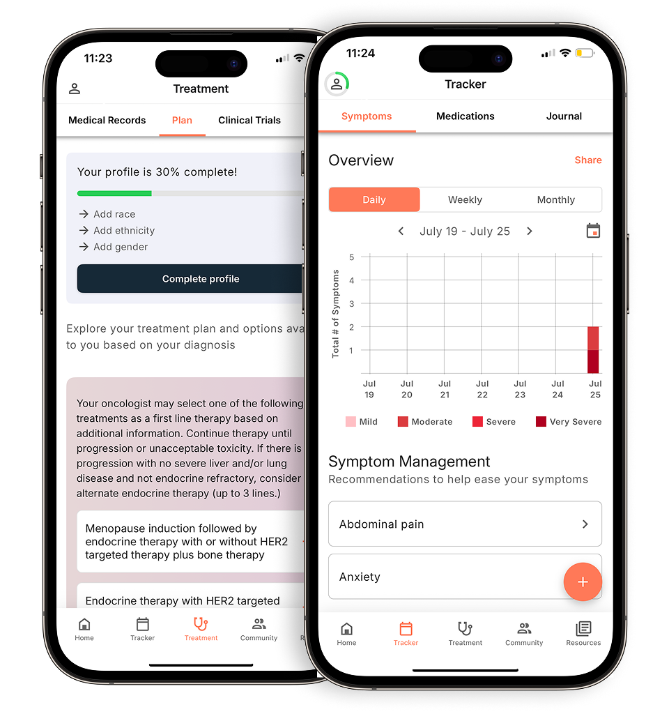
The practical benefit is real. By targeting fewer nodes, surgeons can still get the staging information they need while dramatically reducing the risk of lymphedema and other surgical complications. Patients who are weighing their options before surgery often find this distinction worth discussing in depth with their care team.
Once staging is complete, the pathology results inform the next critical decision: whether additional treatment is needed — and that’s where radiation therapy enters the picture.
Deciding on radiation: the role of the ‘cleanup crew’
After endometrial cancer surgery removes the uterus and nearby tissue, radiation steps in as the cleanup crew — targeting any microscopic cancer cells that may have been left behind. Adjuvant radiation doesn’t treat visible disease; it’s designed to reduce the risk of cancer returning in the pelvis. According to the National Cancer Institute, while radiation after surgery can reduce the risk of cancer returning in the pelvic region, chemotherapy alone after surgery may be equally effective at preventing overall recurrence in women with locally advanced endometrial cancer.
There are two primary delivery methods, and they work very differently:
- External Beam Radiation Therapy (EBRT) directs high-energy X-rays at the entire pelvic region from outside the body. It’s typically reserved for higher-risk cases where cancer cells may have spread to lymph nodes or surrounding tissue. Radiation therapy may cause urinary side effects, including bladder irritation and increased urinary frequency, particularly in patients with baseline urinary symptoms.
- Brachytherapy (internal radiation) places a small radioactive device directly inside the vagina, targeting the vaginal cuff — the surgical site where the uterus was removed. It delivers a concentrated dose with far fewer systemic side effects, making it the preferred option for many lower-risk patients, as noted by the Cancer Council Australia.
Timing matters, too. Radiation typically begins within six to eight weeks after surgery, once the body has had adequate time to heal. Some patients receive EBRT and brachytherapy in combination, while others may need only one — or neither. Which approach is right for you depends heavily on what the pathology report reveals about the tumor itself, a topic the next section explores in detail.
How your pathology report dictates your radiation plan
Your pathology report isn’t just a summary of findings — it’s the document that determines whether you’ll need radiation, and what kind.
Two findings carry the most weight. Tumor grade is part of your pathology report and helps classify your endometrial cancer, with Grade 3 representing high-grade tumors that are considered more serious. Myometrial invasion measures how deeply the tumor grew into the uterine wall — and this is a primary radiation trigger. According to research published in PMC NIH, depth of invasion and tumor grade together are the central factors in deciding whether adjuvant radiation is warranted.
Here’s how those findings shape the decision:
Cancer care guidance for every step of your journey
Get treatment options, clinical trials, and support tailored to your diagnosis--all in one place.
Get started
See treatment options, manage symptoms, and stay informed—all in the app
View treatment options and trials personalized to your diagnosis—plus track progress in real time.
Continue in app- Low-grade + superficial invasion: Patients with Stage 1, Grade 1 tumors and less than 50% myometrial invasion are often considered low-risk. Many require no radiation at all after surgery.
- Intermediate risk: Deeper invasion or a higher grade shifts patients toward adjuvant treatment — typically brachytherapy, which delivers radiation locally to the vaginal cuff.
- High-grade + deep invasion: This profile generally prompts a discussion of brachytherapy vs external beam radiation therapy, since the risk of regional spread is higher and a wider treatment field may be necessary.
Bold callout: For many Stage 1 patients, the pathology report is what keeps them off the radiation table entirely. As Cancer Research UK notes, low-risk Stage 1 disease often doesn’t require radiation after a hysterectomy.
Understanding these numbers — your grade, your invasion depth, your stage — puts you in a much stronger position to ask the right questions. And as you’ll see in the next section, newer genetic markers are adding another layer to this picture entirely.
The future of care: genetic testing and clinical trials
Genetic testing is reshaping how oncologists approach endometrial cancer — moving treatment decisions beyond stage and grade into the molecular blueprint of the tumor itself.
Once your pathology report confirms tumor grade and confirms whether sentinel node mapping uterine cancer uterine cancer staging was performed, the next frontier is molecular profiling. Two markers in particular are changing the treatment landscape:
- MSI-High (microsatellite instability-high) status signals that the tumor has defects in its DNA repair system, making it highly responsive to immunotherapy drugs called checkpoint inhibitors.
- Lynch Syndrome — a hereditary condition linked to approximately 3% of endometrial cancers — doesn’t just affect your treatment plan; it has direct implications for your biological family members’ cancer risk.
- POLE mutations represent an ultra-mutated tumor profile that often carries a surprisingly favorable prognosis, and may reduce the need for aggressive adjuvant therapy.
For patients with MSI-High or mismatch repair-deficient (dMMR) tumors, FDA-approved immunotherapies such as dostarlimab and pembrolizumab are now available treatment options, with some immunotherapy combinations approved for advanced or recurrent endometrial cancer.
Clinical trials, meanwhile, have shifted from a “nothing else worked” option to a legitimate path toward precision medicine. Research shows that personalized treatment approaches including access to clinical trials and targeted therapies selected through genetic testing can offer patients more tailored options for their specific tumor characteristics.
Understanding your molecular profile opens doors your standard pathology report can’t. Knowing which questions to ask your oncology team is the practical next step.
What to ask your oncology team today
Asking the right questions at your next appointment is one of the most practical steps you can take toward personalized care. Compass Oncology recommends that patients take an active role in their cancer care by keeping organized notes, asking questions at appointments, and communicating regularly with their care team.
The questions below are designed to move the conversation from general to specific, helping you confirm that your treatment plan reflects everything your pathology report revealed.
- “What exact grade and stage did surgery confirm?” Your plan should be built on surgical findings, not pre-operative estimates alone.
- “Was sentinel node mapping uterine cancer performed, and what did it show?” Lymph node status directly shapes radiation decisions. If you want to understand more about what that procedure involves, this overview of what to expect can help you frame the conversation.
- “What are the long-term side effects of EBRT compared to brachytherapy for my specific situation?” Side effect profiles differ meaningfully between the two approaches, and your quality-of-life priorities matter here.
- Has molecular profiling or genetic testing been done on my tumor? Your pathology report includes detailed analysis of your tumor tissue, which helps your care team understand your diagnosis and plan your treatment.
Bringing these questions to your appointment doesn’t require medical expertise — it requires preparation. The more precisely your team can answer them, the more confident you can feel that your care is tailored to your specific pathology rather than a generalized protocol. That confidence, combined with the right tools to track your progress, is the foundation of everything covered in the final section ahead.
Navigating your path forward
Understanding your endometrial cancer pathology report isn’t just a medical formality — it’s the foundation every treatment decision is built on, from the operating room through radiation and beyond.
The key insights from this article are worth holding onto:
- Surgery comes first and defines everything else. Your surgical findings — including lymph node status, tumor grade, and depth of myometrial invasion — become the roadmap for all follow-up care.
- Radiation is personalized, not automatic. Whether you receive external beam radiation, vaginal brachytherapy, or no radiation at all depends entirely on what your surgical pathology reveals about your risk level.
- Genetic markers open doors. Mutations like MMR deficiency and POLE status aren’t just diagnostic details — they’re the keys to immunotherapy eligibility and clinical trial matching, which can meaningfully expand your options.
- Specialized pathologists and radiologists can analyze your pathology results to tailor a personalized uterine cancer treatment plan for you, ensuring your care is informed by comprehensive diagnostic testing.
You don’t have to parse this alone. Whether you’re weighing surgical approaches or trying to understand what lymph node results mean for your next steps, having the right tools and questions makes a real difference. Download the Outcomes4Me app to get personalized, evidence-based guidance — and take control of your care.
Disclaimer: The information provided in this article is for informational purposes only and is not intended to be a substitute for professional medical advice, diagnosis, or treatment. Outcomes4Me is not acting as your caregiver, and any suggestions or guidance offered should not replace the advice of your healthcare provider or qualified medical professional. Always seek the guidance of your physician or other qualified health provider with any questions you may have regarding a medical condition.
Personalized support for real care decisions
Understand your diagnosis, explore clinical trials, and track symptoms--all in one place.
Get started
Compare treatments, prepare for appointments, and track side effects—all in the app
Built for your diagnosis, Outcomes4Me gives you the tools to make confident, informed decisions—right when you need them.
Continue in app