
Learning how to read a prostate biopsy pathology report starts at the top, not at the diagnosis line. The header carries your name, date of birth, medical record number, and the ordering provider. Verify every identifier before proceeding. Beneath it sits the gross description, the pathologist’s account of what physically arrived in the lab. How many tissue cylinders, how long each measured, and which container they came from.
Each prostate biopsy core sample is a thin, cylindrical section of tissue retrieved with a spring-propelled needle. Specimen labels such as “Right Base” or “Left Apex” map each core back to a precise location in the gland, and a standard sampling of 12 or more cores determines how thoroughly the gland was searched.
Benign, suspicious, and malignant results
Benign prostatic tissue means normal glandular architecture, often reflecting benign prostatic hyperplasia. Inflammation, or prostatitis, may be noted even when it needs no treatment, and it can also elevate PSA. A prostate biopsy negative for cancer, coupled with a persistently rising PSA, prompts continued surveillance, imaging, or repeat sampling.
Suspicious findings. Understanding PIN and ASAP
Prostatic intraepithelial neoplasia describes abnormal cells confined within existing glands. Low-grade PIN carries little clinical weight; high-grade PIN (HGPIN), particularly when multifocal, warrants closer follow-up. Atypical small acinar proliferation (ASAP) is a borderline call, cells suspicious for cancer but too few or too distorted to confirm. These atypical prostate biopsy results are not malignancies, yet ASAP frequently triggers a repeat biopsy.
The Gleason score and grade group systems
When cancer is present, the report names it: adenocarcinoma, meaning a malignancy arising from the gland-forming cells that make up prostate tissue. The pathologist then grades it by architecture, not by the individual appearance of the cells.
Pattern 3 describes well-differentiated glands that remain separate and recognizable. Pattern 4 shows glands fusing, losing definition, or forming sieve-like structures. Pattern 5 shows sheets or cords of cells with no gland formation at all. The prostate biopsy Gleason score combines the most common pattern with the second most common.
Because the older scale effectively began at 6, the ISUP grade group prostate system reframes prostate biopsy cancer grades as 1 through 5, with Grade Group 1 the least aggressive. Higher grades correlate with a greater likelihood that disease extends beyond the capsule.
Evidence-based guidance powered by NCCN Guidelines®
Personalized treatment plans shaped by the latest oncology standards—tailored to your diagnosis.
Get started
View your personalized treatment plan in the Outcomes4Me app
Use your diagnosis to unlock personalized NCCN Guidelines®-aligned recommendations.
Continue in app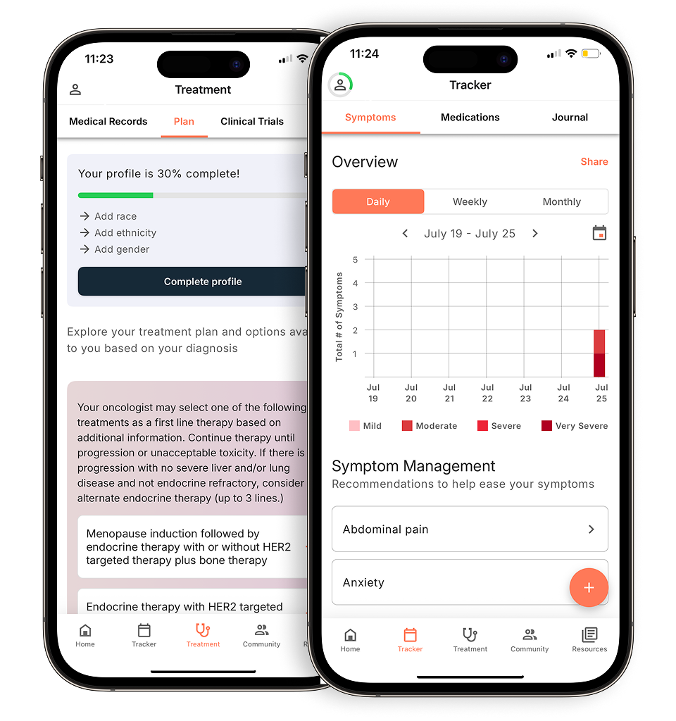
Calculating the Gleason sum: 3+4 vs. 4+3
The first number is the dominant pattern. What does Gleason 7 mean? It depends on order. In the Gleason 3+4 vs 4+3 distinction, 3+4 (Grade Group 2) is mostly well-formed glands with a minority of pattern 4. 4+3 (Grade Group 3) is predominantly pattern 4. That reversal typically moves treatment planning from possible surveillance toward definitive therapy.
Quantifying the Cancer. Tumor volume and location cata
Grade answers how aggressive. Volume answers how much. Your report lists, core by core, the percentage of cancer in prostate biopsy tissue, the fraction of that specific cylinder replaced by tumor.
Your pathology report often includes how many biopsy cores contain cancer, which shows whether cancer is present in different areas of the prostate. Two positive cores out of twelve describe a very different clinical situation than ten.
Location matters just as much. Cores are labeled by side and by zone, so the pattern of positivity tells your care team whether disease appears unilateral, confined to one region, or bilateral and diffuse. That map may help your surgical team consider nerve-sparing approaches during surgery and may inform dose planning in radiation therapy. Ask your care team how this information factors into their treatment planning for your specific situation.
Perineural invasion may also appear here, cancer cells tracking alongside or encircling the small nerves that run through the gland.
Tumor percentages and linear millimeters
Your pathologist will report findings for each core (biopsy sample) taken from your prostate during the procedure. Both measure volume, but millimeters are less affected by how long the core happened to be. Prostate biopsy cancer in one core at high percentage may represent focal disease, while small amounts scattered across many cores signal multifocal disease, which often disqualifies a patient from focal therapy.
Perineural invasion (PNI) and vascular space invasion
Perineural invasion (PNI) describes cancer invading the space surrounding small nerve fibers within the prostate, and its presence on a biopsy is associated with a greater chance that the cancer may become more aggressive during surveillance or be found outside the prostate. Lymphovascular invasion (LVI), cancer within lymphatic or blood vessel spaces, is a stronger warning because those channels carry cells to distant sites. Both PNI and LVI are microscopic findings that your doctor may consider as part of overall risk assessment when planning next steps, which may or may not include imaging scans.
Cancer care guidance for every step of your journey
Get treatment options, clinical trials, and support tailored to your diagnosis--all in one place.
Get started
See treatment options, manage symptoms, and stay informed—all in the app
View treatment options and trials personalized to your diagnosis—plus track progress in real time.
Continue in appThe role of immunohistochemistry (IHC) stains
Some slides resist a confident call under standard hematoxylin and eosin staining. When the pathologist applies immunohistochemistry, antibody-based dyes bind to specific proteins in the tissue sample. This staining technique can help highlight certain characteristics of the glands that may be useful in evaluating your biopsy.
Three prostate biopsy pathology abbreviations dominate this part of the report. P63 and high molecular weight cytokeratin (HMWCK, sometimes listed as 34βE12) mark basal cells. AMACR, also written P504S, marks a tumor-associated enzyme.
Most labs run these together as a “cocktail.” The absence of basal cell markers combined with positive AMACR staining may support a diagnosis of adenocarcinoma, though your pathologist will interpret these findings together with other features of the tissue sample. Your pathologist may use special stains to look for basal cells, which can help distinguish benign prostate glands from cancer cells.
These stains are the accepted method for resolving suspicious and ASAP findings, and they belong in your understanding of prostate biopsy pathology terms.
What the report doesn’t tell you
Reading prostate biopsy results well means knowing what the document cannot say.
Template biopsy takes only a small number of tissue samples from the prostate, meaning the biopsy may not represent the entire gland, particularly in larger prostates. A clinically significant lesion can sometimes sit between needle tracks, which is why your doctor may discuss whether additional imaging or biopsy approaches could help ensure nothing is missed.
Distinguishing between pattern 3 and pattern 4 can be challenging, and pathologists may interpret these boundaries differently from one another.
In about 25 percent of cases, the Gleason score on a biopsy may underestimate the true grade of cancer in the entire prostate, meaning the biopsy may not always reflect the worst disease present.
A separate pathology report will be created if your prostate is removed surgically to treat cancer, and this report may contain different findings than your biopsy report.
Your care team may consider PSA density, MRI PI-RADS scoring, family history, and germline testing as part of understanding your overall risk profile. Ask your doctor which of these factors apply to your situation.
The need for a second opinion
Slides and blocks belong to you, and you can request them from the hospital where your biopsy was performed to send to another doctor or institution for review. Understanding your biopsy pathology report is strongest when the grading is confirmed. Review matters most at decision thresholds, Grade Group 1 versus 2, 3+4 versus 4+3, and for rare variants. Ask specifically for an expert urologic pathologist.
Common misconceptions about biopsy accuracy
A negative biopsy result does not rule out prostate cancer, which is why PSA monitoring continues. When prostate biopsy results explained by grade alone leave treatment ambiguous, genomic classifiers such as Decipher or Prolaris can add independent risk information from the same tissue.
Key Takeaways: What You Need to Know
- Grade drives decisions. The Gleason score and ISUP Grade Group influence treatment recommendations more than any other single element of the report.
- Read the comment section. Pathologists reserve their nuance in the comment section, diagnostic uncertainty, cribriform architecture, IHC reasoning, and recommendations for repeat sampling.
- Bring the full document. The pathologist gives each biopsy core its own diagnosis in your full pathology report, so it’s important to review the diagnoses for all of the cores to understand your results.
- Treat HGPIN and ASAP as action items. No cancer was found with HGPIN and ASAP findings, but these findings call for a defined follow-up schedule rather than passive waiting.
- Track your numbers over time. Logging PSA values, imaging, and prostate biopsy report examples from each round keeps the full record available when you discuss a personalized care plan with your care team.
Disclaimer: The information provided in this article is for informational purposes only and is not intended to be a substitute for professional medical advice, diagnosis, or treatment. Outcomes4Me is not acting as your caregiver, and any suggestions or guidance offered should not replace the advice of your healthcare provider or qualified medical professional. Always seek the guidance of your physician or other qualified health provider with any questions you may have regarding a medical condition.
Personalized support for real care decisions
Understand your diagnosis, explore clinical trials, and track symptoms--all in one place.
Get started
Compare treatments, prepare for appointments, and track side effects—all in the app
Built for your diagnosis, Outcomes4Me gives you the tools to make confident, informed decisions—right when you need them.
Continue in app