
Understanding how metastatic colorectal cancer (mCRC) spreads can help you ask informed questions, better understand your options, and take a more active role in your care.
mCRC occurs when cancer cells break away from the original tumor in the colon or rectum and spread to distant parts of the body. Awareness around mCRC has grown significantly, and for good reason: CRC remains one of the most common cancers, and many patients are diagnosed after the disease has already spread.
CRC frequently spreads to the liver, which comes down to anatomy. Blood from the intestines drains directly into the portal vein, which carries blood to the liver. This creates a biological pathway that allows circulating cancer cells from the colon to reach the liver early in the metastatic process.
Research also suggests that metastatic spread often begins before the primary tumor is detected. Several genomic sequencing studies, including research from Stanford University, have shown that microscopic metastatic “seeding” can occur years before diagnosis in some patients. Beyond the liver, colorectal cancer may also spread to the lungs, lymph nodes, or the lining of the abdominal cavity, known as the peritoneum.
Evidence-based guidance powered by NCCN Guidelines®
Personalized treatment plans shaped by the latest oncology standards—tailored to your diagnosis.
Get started
View your personalized treatment plan in the Outcomes4Me app
Use your diagnosis to unlock personalized NCCN Guidelines®-aligned recommendations.
Continue in app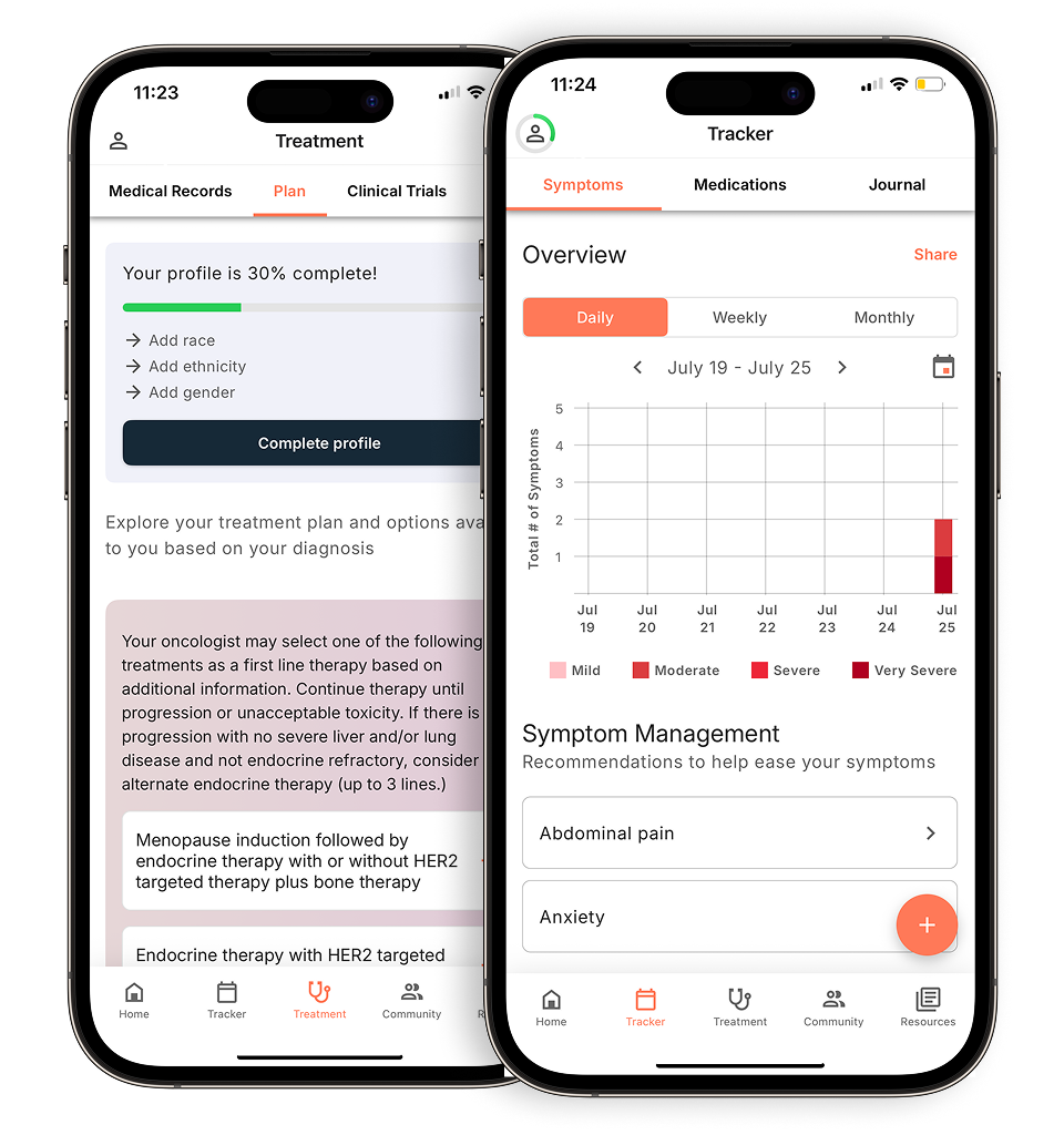
The liver: The most common site of spread
Studies estimate that up to 60% of patients with CRC will develop liver metastases at some point during their disease course.
Not all liver metastases are the same.
- Synchronous metastases are present at the time of the initial cancer diagnosis.
- Metachronous metastases develop later, sometimes months or years after treatment of the primary tumor.
This distinction matters because some patients with liver-only disease may be eligible for surgery or liver-directed therapies with curative intent. However, eligibility depends on factors such as the number and size of tumors, their location, and overall liver function.
While liver involvement is most common, rectal cancer can follow a different metastatic pattern.
The lungs and brain: Why rectal cancer can spread differently
Rectal cancer doesn’t always spread in the same way as colon cancer, although the liver is still a common site. Some rectal cancer cells can travel directly to the lungs.
Lung metastases occur more frequently in rectal cancer than in colon cancer, which is why chest imaging is particularly important during surveillance for many rectal cancer patients.
Key differences for rectal cancer patients to know include:
- Higher risk of lung metastases: Rectal cancer spreads to the lungs more often than colon cancer.
- Different surveillance needs: Chest CT scans may play a larger role in follow-up care.
- Brain metastases are uncommon: Brain involvement is relatively rare in colorectal cancer overall, but the risk increases in advanced systemic disease.
Because metastatic patterns vary depending on where the cancer started, follow-up imaging is often individualized.
Peritoneal metastases: A more difficult form of spread to detect
CRC can also spread to the peritoneum, the thin lining surrounding the abdominal organs. This is known as peritoneal metastasis or peritoneal carcinomatosis.
Peritoneal spread can be especially challenging to detect because tumors may appear as tiny, diffuse deposits rather than large, clearly defined masses on imaging scans.
Several factors contribute to this difficulty:
- Small tumor implants may fall below the resolution of standard imaging
- Disease can spread diffusely across the abdominal lining
- Symptoms are often vague, including bloating, abdominal discomfort, or changes in appetite
Peritoneal metastases are classified as M1c disease and are generally associated with a poorer prognosis compared with isolated liver or lung metastases.
For carefully selected patients, treatment options may include cytoreductive surgery combined with hyperthermic intraperitoneal chemotherapy (HIPEC), which delivers heated chemotherapy directly into the abdominal cavity during surgery.
Why surveillance and biomarker testing matter
Surveillance after a CRC diagnosis is not one-size-fits-all.
The location of the primary tumor, extent of disease, and tumor biology all help determine how patients are monitored over time. For example, patients with rectal cancer may need closer chest surveillance because of the increased risk of lung metastases.
Biomarker testing can also provide important information. Mutations such as KRAS, NRAS, and BRAF influence treatment decisions and may also correlate with differences in disease behavior and prognosis.
Questions worth asking your care team include:
- Why were these imaging intervals chosen for me?
- Should I have routine chest imaging?
- Have I received comprehensive biomarker testing?
- How do my biomarker results affect my treatment options?
Understanding the reasoning behind your surveillance plan can help you feel more informed. mCRC is increasingly managed as a biologically complex disease, not a single uniform diagnosis. Understanding your cancer’s specific characteristics can help guide more personalized treatment decisions and conversations with your care team.
Connect with an Outcomes4Me oncology nurse practitioner at no charge through the Outcomes4Me app, using the “Ask Outcomes4Me” button.
Personalized support for real care decisions
Understand your diagnosis, explore clinical trials, and track symptoms--all in one place.
Get started
Compare treatments, prepare for appointments, and track side effects—all in the app
Built for your diagnosis, Outcomes4Me gives you the tools to make confident, informed decisions—right when you need them.
Continue in app