
The fundamentals of lung cancer lymph node involvement
When a pathology report mentions the lymph nodes, understanding what that finding actually means helps you prepare for what comes next.
Your care team uses TNM staging, which includes Tumor size, Node involvement, and Metastasis spread, to describe how far lung cancer has developed. The “N” component specifically maps whether cancer cells have reached the regional lymph nodes, the small immune-filtering stations positioned throughout the chest that process fluid draining from lung tissue. Their location makes them a natural checkpoint where cancer cells may travel before spreading further.
One point worth understanding early: lymph node enlargement doesn’t automatically mean cancer has spread. Nodes can swell in response to infection or inflammation, which is why your care team confirms involvement through imaging and, often, biopsy. And while lung cancer can present differently across individuals, some people notice symptoms like persistent fatigue or mild shortness of breath rather than a classic cough, N-staging focuses on the anatomical spread of cancer, not symptoms.
Understanding N-staging classifications
When early signs of lung cancer symptoms prompt imaging, the resulting scans map not just the tumor but the surrounding lymph node stations. N0 means no detectable nodal involvement. N1 indicates spread to nodes inside the lung or at the hilum, the point where the airway meets the lung tissue. N2 signals involvement of mediastinal nodes on the same side as the primary tumor, while N3 covers distant nodes such as those in the neck (supraclavicular) or on the opposite side of the chest. Each step outward represents a wider anatomical reach, which directly shapes the treatment options your care team will consider.
How confidently those classifications are assigned depends on whether staging is based on imaging alone or confirmed tissue, a distinction the next section unpacks in full. To learn more about non-small cell lung cancer staging and prognosis, your care team can help you understand what each stage classification means for your specific situation.
Clinical vs. pathologic staging. Why the numbers might change
Your N-stage may change as your care team gathers more information. Imaging gives your care team a working map, but surgical biopsy often provides more precise findings.
Clinical staging (cTNM) relies on CT scans, PET scans, and other imaging to assess nodal involvement before any tissue is removed. It’s the first picture your care team builds, often prompted by lung cancer symptoms like a persistent cough, chest pain, or unexplained fatigue. That picture guides initial treatment decisions but carries inherent limits, as imaging can miss microscopic nodal disease or flag inflammation as cancer.
Pathologic staging (pTNM) is based on direct microscopic examination of tissue collected during surgery or biopsy. Because it analyzes actual cells rather than indirect signals, it’s considered the gold standard for accuracy. A node that appeared clean on a PET scan may reveal micrometastases under a microscope, or a suspicious node may turn out to be benign.
This gap between the two methods means stage shifting is a real possibility. After surgical pathology results return, your N-classification may move up or down. That reflects how diagnostic precision deepens over time. Understanding this helps set realistic expectations as your personalized care plan takes shape. And as the next section explores, knowing where cancer has spread anatomically is only part of the picture, how aggressively it behaves biologically is a separate, equally important question.
Evidence-based guidance powered by NCCN Guidelines®
Personalized treatment plans shaped by the latest oncology standards—tailored to your diagnosis.
Get started
View your personalized treatment plan in the Outcomes4Me app
Use your diagnosis to unlock personalized NCCN Guidelines®-aligned recommendations.
Continue in app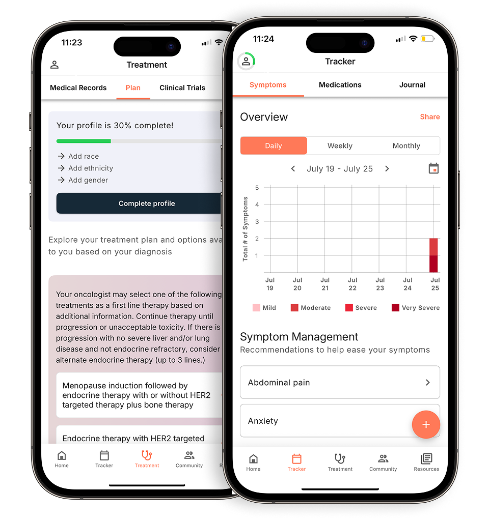
Conventional wisdom vs. reality: the lymph node aggression myth
When people hear that lung cancer has reached the lymph nodes, a common fear is that the disease is inherently aggressive or beyond meaningful lung cancer treatment. That concern is understandable, but it conflates two separate questions. Where the cancer is and how it behaves biologically. The N-classification tells your care team which nodal stations are involved, not the tumor’s growth rate.
Biology often tells the more important story. Mutations such as EGFR or ALK can make a cancer highly responsive to targeted therapies regardless of its N-stage. A tumor with N2 nodal involvement but a targetable mutation may respond better to treatment than an N1 tumor without one. Location and biology are distinct variables, and your care team evaluates both.
It’s also worth noting that N1 and N3 involvement carry meaningfully different implications for surgical options. N1 disease frequently remains operable, while N3 involvement shifts the focus toward systemic approaches. That distinction is about treatment planning. And as the next section explores, exactly where those nodes are located drives specific, well-defined decisions about your personalized care plan.
How lymph node location dictates your treatment path
Where the affected nodes sit on the anatomical map directly determines whether surgery, systemic therapy, or a combination leads your personalized care plan.
N1 involvement means cancer has spread to lymph nodes within the lung and/or around the area where the bronchus enters the lung, and patients with N1 disease may be considered for Stage 2A or 2B lung cancer depending on tumor size. For locally advanced lung cancer with N2 disease, treatment often involves a combination of chemotherapy, radiation therapy, and sometimes surgery. Immunotherapy may also be used, particularly durvalumab after chemotherapy and radiation are complete for patients who cannot have surgery. When lung cancer reaches N3 lymph nodes (contralateral or supraclavicular nodes), chemotherapy combined with radiation therapy is typically the main treatment approach rather than surgery alone. This anatomical threshold is what clinicians mean when they describe a tumor as “unresectable.”
The role of mediastinoscopy and EBUS in staging
Before your care team can finalize a personalized care plan, they need tissue-confirmed nodal mapping, not just imaging estimates.
Endobronchial ultrasound (EBUS) is now the standard first-line tool for sampling suspicious mediastinal nodes. A bronchoscope with an ultrasound tip is guided into the airways, allowing real-time needle biopsies of nodes that imaging flagged, all without open surgery. When asking what are symptoms for lung cancer that prompted a scan in the first place, it’s worth knowing that EBUS is often what confirms whether nodes are truly involved. Mediastinoscopy is another procedure that can be used for pre-treatment staging to examine whether lymph nodes in the chest are cancerous, as an alternative to EBUS-TBNA. Accurate nodal mapping before radiation therapy is especially critical, as misidentifying involved nodes can leave disease untreated while exposing healthy tissue to unnecessary dose.
Cancer care guidance for every step of your journey
Get treatment options, clinical trials, and support tailored to your diagnosis--all in one place.
Get started
See treatment options, manage symptoms, and stay informed—all in the app
View treatment options and trials personalized to your diagnosis—plus track progress in real time.
Continue in appPrognosis and life expectancy: interpreting the data
N-staging informs how your care team reads the statistical landscape. It shapes how oncologists interpret survival data, but biology, treatment response, and overall health all shift the picture meaningfully.
When people ask what life expectancy looks like once lung cancer involves the lymph nodes, the honest answer is it varies considerably, and the N-stage is only one input. In 2026, immunotherapy protocols have reshaped outcomes for N2 and N3 patients who once had limited options, as combination approaches can achieve durable responses in cases previously considered difficult to manage.
Not all N2 disease carries identical weight, either. Skip metastasis, where a node is involved without sequential spread through adjacent stations, may have different prognostic implications than bulky N2 disease, where multiple large nodes are affected. Your care team can discuss how these patterns might relate to your specific situation.
It’s also worth knowing that N3 involvement, which can extend to the lymph nodes of the neck (supraclavicular region), marks a boundary where systemic and combined-modality strategies take precedence. That shapes how aggressively treatment is pursued. As the next section explores, understanding how quickly lung cancer can progress between stages adds another layer to this picture.
Stage 1 to 4: how quickly does lung cancer spread?
How fast lung cancer moves from an early stage to advanced disease depends heavily on cell type, biology, and individual risk factors. There’s no fixed timeline for progression.
Lung cancer and symptoms often appear gradually, which is part of what makes early detection so difficult. Small cell lung cancer (SCLC) tends to grow and spread more quickly than other types of lung cancer, and patients may progress from localized to more widespread disease relatively rapidly. Non-small cell lung cancer (NSCLC) progression varies widely from person to person, and some patients may experience a slower progression before reaching advanced stages. That fundamental difference changes how urgently treatment begins. Factors like active smoking status, specific genetic markers, and tumor histology all influence the pace. Understanding spread as non-linear, rather than a predictable countdown, is essential context before reading survival benchmarks by N-stage, which the next section addresses directly.
Limitations and considerations in N-staging
N-staging gives your care team a working map, not a complete biological portrait. Understanding where that map has edges helps you ask better questions.
For both non-small cell and small cell lung cancer, the staging picture carries real constraints worth knowing. PET scan false positives are a genuine limitation. Inflammation from a recent infection or pneumonia can cause lymph nodes to light up on imaging in ways that mimic malignant involvement. That’s not a flaw in your scan, it’s a physiological reality that tissue confirmation is designed to catch.
Occult nodal involvement adds another layer of complexity. Nodes that appear entirely normal on imaging can harbor microscopic cancer cells undetectable by any current scan. This is one reason pathologic staging, confirmed at surgery, sometimes reveals more extensive disease than clinical staging suggested.
And even when staging is accurate at the time of diagnosis, it’s a snapshot. Tumor biology evolves, and a staging classification assigned on one date doesn’t fully capture how aggressively the disease may behave over the following months. For complex N2 cases especially, a multidisciplinary tumor board pulling together thoracic surgeons, oncologists, radiation specialists, and pathologists is best positioned to interpret ambiguous findings and align on a personalized care plan. These are precisely the scenarios where staging errors can occur, a dynamic the next section examines directly.
Common questions about lung cancer and lymph nodes
Four questions come up repeatedly when patients receive a lymph node-positive diagnosis, and clear answers help you engage your care team with confidence.
Pain and lymph nodes don’t always go together the way you might expect. Involved nodes rarely cause obvious discomfort on their own. When pain does occur, it’s typically because an enlarged node is pressing on a nearby structure, a nerve, airway, or blood vessel, rather than the cancer itself signaling its presence. That distinction matters when you’re trying to interpret new symptoms between appointments.
Nodal response to treatment is a realistic outcome. After chemotherapy, immunotherapy, or radiation, imaging often shows meaningful shrinkage in previously enlarged nodes, a response that can shift the staging picture and open up options that weren’t available at diagnosis.
Mediastinal and lymph node anatomy is a distinction your care team tracks closely. Mediastinal nodes occupy the central chest area between the lungs. Lymph nodes in these regions are important to evaluate during lung cancer staging. Whether you’re dealing with non-small cell or small cell lung cancer, sometimes called oat cell carcinoma, that anatomical location drives treatment direction.
The disease name doesn’t change with nodal involvement. When cancer spreads to lymph nodes far from the original tumor, it is still classified as lung cancer because the cancer type is determined by where it originated in the body.
What you need to know: key takeaways
N-staging is the clinical framework that tells you, and your care team, exactly where lymph nodes are involved and how far the cancer has spread. That anatomical precision determines everything from whether surgery is on the table to which systemic therapies your personalized care plan will include.
A few principles are worth holding onto as you move forward:
- Pathologic staging beats imaging. Biopsy confirmation via EBUS or surgical sampling is more accurate than CT or PET alone, and the distinction matters when treatment decisions hinge on N1 versus N2 classification.
- N-stage reshapes treatment, not just prognosis. Each step from N1 to N3 shifts the approach, from resection to multimodality or fully systemic care.
- Tumor genetics now share the stage with anatomy. In 2026, your tumor’s molecular profile increasingly drives prognosis alongside N-classification.
- Nodal involvement is treatable in many cases. Many N1 and even N2 cases can be managed with curative intent.
Disclaimer: The information provided in this article is for informational purposes only and is not intended to be a substitute for professional medical advice, diagnosis, or treatment. Outcomes4Me is not acting as your caregiver, and any suggestions or guidance offered should not replace the advice of your healthcare provider or qualified medical professional. Always seek the guidance of your physician or other qualified health provider with any questions you may have regarding a medical condition.
Personalized support for real care decisions
Understand your diagnosis, explore clinical trials, and track symptoms--all in one place.
Get started
Compare treatments, prepare for appointments, and track side effects—all in the app
Built for your diagnosis, Outcomes4Me gives you the tools to make confident, informed decisions—right when you need them.
Continue in app