
The reality of RCC recurrence: why your follow-up plan is a lifeline
Finishing treatment for renal cell carcinoma (RCC) brings real relief, but for many patients, it also brings a quieter, persistent worry: what if it comes back? That fear is valid, and it deserves a direct, honest answer.
Structured surveillance after kidney cancer treatment is a proactive, evidence-based strategy designed to catch problems early, when intervention is most effective.
Most recurrences occur within the first three to five years after surgery. That window calls for a plan. Recognizing kidney cancer recurrence symptoms early, whether through imaging or a clinical exam, can meaningfully change your options.
Not every patient carries the same risk, which is exactly why a one-size-fits-all schedule doesn’t work. Oncology teams use risk stratification to classify patients as lower risk or higher risk based on tumor stage, grade, and other pathological factors, and determine how frequently you should be scanned and which imaging studies are appropriate. A patient who had a small, low-grade tumor removed with clear margins follows a very different schedule than someone who presented with locally advanced disease.
That’s the foundation of personalized follow-up: your specific biology, your specific surgery, and your specific risk profile all shape the plan. The data your care team collects over time is your best defense. Understanding what to look for between appointments is equally important, and that starts with knowing the difference between local and systemic warning signs.
Recognizing the signs: localized vs. systemic symptoms
Knowing what to watch for between appointments is one of the most practical steps you can take within any RCC follow-up protocol, but it’s equally important to know what those signs actually mean.
Renal cell carcinoma recurrence can appear in two distinct ways: as a local recurrence near the original surgical site, or as metastatic spread to distant organs. Understanding the difference helps you communicate more clearly with your care team when something feels off.
Local recurrence signals are often felt closer to home. Red flags tied to the surgical site include:
- Flank pain or persistent discomfort on the side where your kidney was removed
- Blood in the urine (hematuria)
- A palpable mass or unusual fullness in the abdomen
Systemic symptoms, on the other hand, suggest the cancer may have spread beyond the kidney region. The lungs are the most common site for RCC metastasis, which means pulmonary symptoms deserve particular attention. Watch for:
- A persistent or unexplained cough
- Shortness of breath that isn’t explained by another condition
- Bone pain, which may signal skeletal involvement
Routine imaging identifies most recurrences before you’d ever feel any symptoms. That reality underscores why scheduled scans are the true backbone of post-treatment surveillance. The next section breaks down exactly which tools your care team uses to catch what you can’t feel.
Evidence-based guidance powered by NCCN Guidelines®
Personalized treatment plans shaped by the latest oncology standards—tailored to your diagnosis.
Get started
View your personalized treatment plan in the Outcomes4Me app
Use your diagnosis to unlock personalized NCCN Guidelines®-aligned recommendations.
Continue in app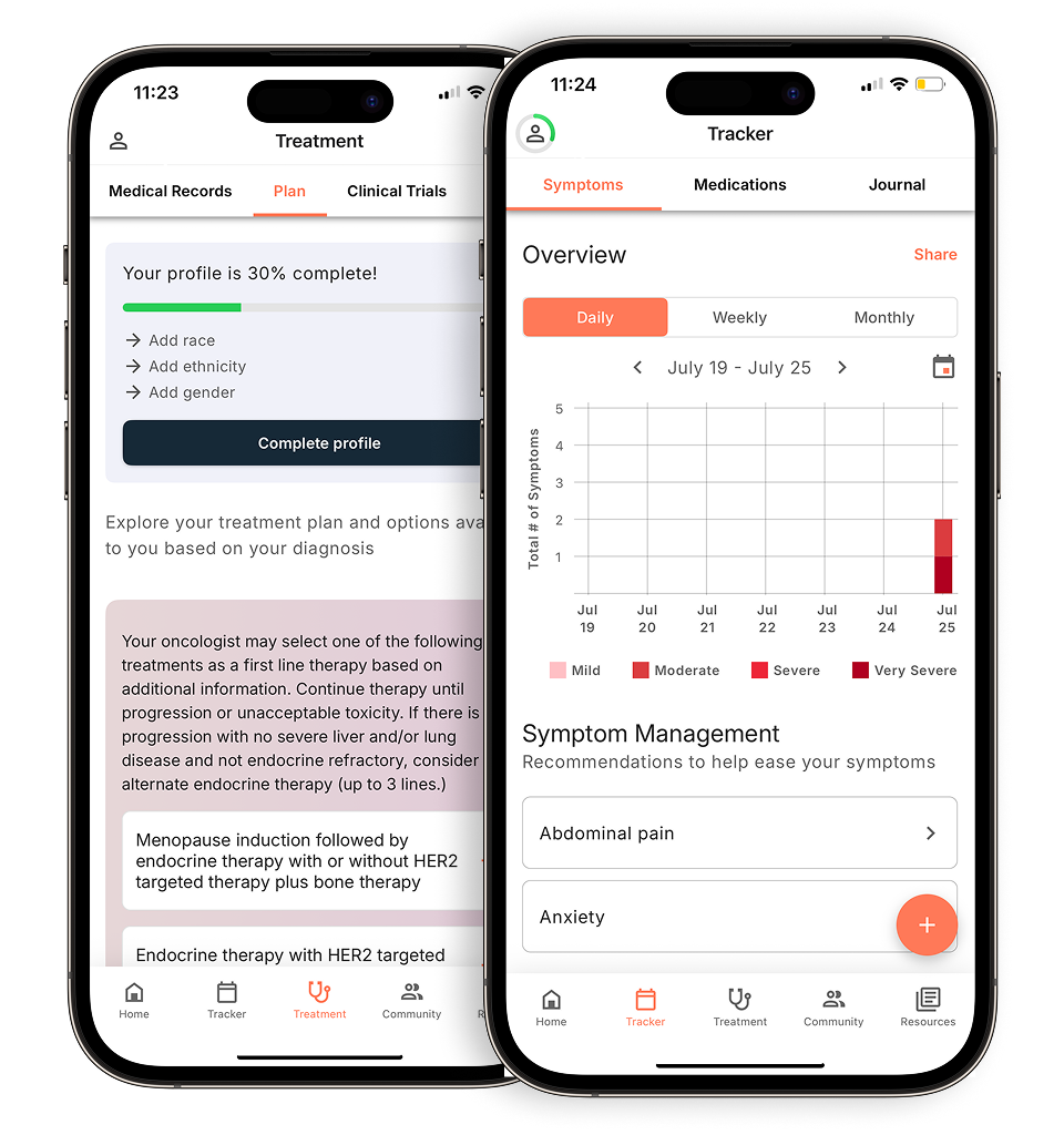
Understanding the surveillance toolkit: scans and blood work
Surveillance after RCC treatment is only as effective as the tools behind it, and knowing what each test is actually looking for makes those follow-up appointments far less intimidating.
Imaging is the backbone of any post-treatment monitoring plan. CT scans of the abdomen and pelvis are the primary method for checking the remaining kidney tissue, the surgical site, and nearby lymph nodes for signs of local recurrence. MRI offers an alternative, particularly useful when contrast dye is a concern or when soft-tissue detail matters most. Life after nephrectomy involves your remaining kidney doing the work of two, so imaging helps catch cancer early and shows how that kidney is functioning under increased demand.
Chest imaging, either X-ray or CT, is non-negotiable. The lungs are the most common site of RCC metastasis, and early detection of pulmonary nodules significantly widens treatment options. Your care team may use chest CT as part of your surveillance plan, as it can help detect small lung lesions that might not be visible on a standard X-ray.
Blood work runs alongside imaging to fill gaps that scans can’t cover. Two markers get particular attention:
- eGFR and creatinine track how well your remaining kidney is filtering waste. A declining eGFR can signal chronic kidney disease developing post-surgery, a common concern that requires its own management plan.
- Calcium and liver function tests (LFTs) serve as indirect red flags. Your care team may monitor blood work such as calcium levels and liver function tests during follow-up, as these can sometimes reflect changes that warrant further investigation.
| Test | Purpose | Typical frequency |
|---|---|---|
| CT abdomen/pelvis | Detect local recurrence or lymph node involvement | Every 6–12 months (varies by risk) |
| Chest CT or X-ray | Screen for pulmonary metastasis | Every 6–12 months |
| eGFR / creatinine | Monitor remaining kidney function | At each follow-up visit |
| Calcium / LFTs | Flag possible bone or liver metastasis | At each follow-up visit |
The frequency of these tests is tied directly to where your cancer was staged at diagnosis. That connection between your pathology results and your surveillance schedule is where the concept of risk stratification becomes central to your care plan.
Risk stratification: how your stage determines your schedule
Your follow-up schedule after RCC treatment is built directly on your pathology results, and understanding those results helps you see why renal cell carcinoma surveillance guidelines are calibrated the way they are.
Cancer care guidance for every step of your journey
Get treatment options, clinical trials, and support tailored to your diagnosis--all in one place.
Get started
See treatment options, manage symptoms, and stay informed—all in the app
View treatment options and trials personalized to your diagnosis—plus track progress in real time.
Continue in appTwo key factors drive your risk profile: TNM stage and Fuhrman grade. TNM describes the tumor’s size and spread, “T” for the primary tumor, “N” for lymph node involvement, “M” for metastasis. Fuhrman grade (or the more current WHO/ISUP grade) reflects how aggressive the cancer cells look under a microscope, rated one through four. A higher T-stage classification (T3 or T4) places patients in a higher risk category for recurrence after kidney cancer treatment.
For patients with low-risk disease, typically T1 tumors with low grades, monitoring tends to be less intensive. According to surveillance guidelines, patients with low-risk disease may have less frequent follow-up visits after the first five years, with imaging and lab work spaced further apart (occurring between 72–120 months and beyond). High-risk patients, however, follow a different path entirely. T3–T4 disease typically calls for imaging every three to six months in the first few years, with ongoing surveillance extending well beyond the five-year mark, and in many cases, lifelong monitoring.
The first three years are the period of greatest vigilance for most patients, since recurrence rates peak within this window regardless of stage. That concentration of appointments can feel relentless, and the emotional weight of that schedule is something worth addressing on its own.
Managing ‘scanxiety’ and the mental burden of survivorship
Surveillance after kidney cancer treatment carries real psychological weight, and it deserves equal attention to the physical process. “Scanxiety,” the spike in anxiety that builds in the days before an imaging appointment, is a standard part of the survivor experience. It doesn’t signal weakness; it signals how much is at stake.
Preparation is one of the most effective antidotes to that anxiety. In practice, patients who arrive at appointments with a written list of specific questions about their imaging results, what abnormal blood work means, or whether symptoms like fatigue or elevated calcium are concerning tend to feel more in control of the conversation. Organized records matter, too. Keeping a running folder of past scans, lab results, and pathology reports reduces the chaos of trying to reconstruct your history at each visit.
A strong support system is most valuable during the three-to-five-year peak window after surgery. Whether that’s a trusted family member who attends appointments, a patient community, or a care navigator, having people who understand your surveillance cadence helps you stay grounded between scans. The next section brings together the most critical things to keep in mind as you move forward in survivorship.
What you need to know: key takeaways for survivors
Understanding your kidney cancer survival rate improves when you stay informed, stay consistent with follow-up, and know exactly what your care team is watching for. The sections above have covered a lot of ground, here’s what matters most to keep close.
Recurrence most often appears in the lungs and the remaining kidney tissue, making chest imaging and renal function monitoring two pillars of any solid surveillance plan. Approximately 20–30% of patients with localized RCC will experience recurrence, which is precisely why consistent follow-up is essential.
- Your stage and grade drive your schedule. A low-grade Stage I tumor carries a very different monitoring rhythm than a high-grade Stage III diagnosis. There’s no universal timeline.
- The first 36 months post-surgery are the highest-risk window. Recurrence is most likely to emerge early, so imaging frequency is deliberately front-loaded during this period.
- Blood tests are early warning tools. Calcium and liver function panels can flag metabolic changes before symptoms appear, giving your team a critical head start.
- Missing a scan has real consequences. Even a single skipped imaging appointment can delay detection by months, time that matters enormously in a recurrence scenario.
Staying on top of these fundamentals puts you in a stronger position than passive waiting ever could. The next step is working with your care team to ensure your surveillance plan matches your individual risk profile and circumstances.
Taking control: your role in personalized survivorship
Staying informed is one of the most powerful things you can do after kidney cancer treatment, and having the right tools makes that possible. Some digital platforms are designed to help patients align their care with established treatment guidelines like those from the National Comprehensive Cancer Network (NCCN®). Ask your care team whether tools like these might support your personalized surveillance plan.
Evidence-based survivorship requires consistent engagement: attending follow-up appointments, understanding what your imaging results mean, knowing when something has changed. The surveillance toolkit, your imaging schedule, lab work, and specialist visits, is most effective when you’re an active participant in it, not just a recipient of appointments.
Every follow-up scan, every conversation with your provider, and every informed question you ask contributes to better outcomes. Work with your care team to build a surveillance plan that reflects your specific situation, and stay engaged with the evidence and tools that support your long-term health.
Disclaimer: The information provided in this article is for informational purposes only and is not intended to be a substitute for professional medical advice, diagnosis, or treatment. Outcomes4Me is not acting as your caregiver, and any suggestions or guidance offered should not replace the advice of your healthcare provider or qualified medical professional. Always seek the guidance of your physician or other qualified health provider with any questions you may have regarding a medical condition.
Personalized support for real care decisions
Understand your diagnosis, explore clinical trials, and track symptoms--all in one place.
Get started
Compare treatments, prepare for appointments, and track side effects—all in the app
Built for your diagnosis, Outcomes4Me gives you the tools to make confident, informed decisions—right when you need them.
Continue in app