
The diagnostic path: understanding papillary renal cell carcinoma subtypes
Whether a papillary renal cell carcinoma diagnosis is Type 1 or Type 2 shapes what follows
Renal cell carcinoma (RCC) is a category of diseases rather than a single disease, and papillary RCC is its second most common form. Papillary renal cell cancer is the second most common subtype, representing roughly 10% to 15% of all kidney cancers. Within that category, Type 1 and Type 2 are biologically distinct, with differences visible at the cellular level. Type 1 papillary renal cell carcinoma is associated with activation of the MET oncogene, which distinguishes it from type 2 papillary RCC. Type 2 papillary renal cell carcinoma has distinct morphological features that differ from type 1, though recent research suggests these differences may represent morphologic variation rather than a separate disease category.
For most patients, the path starts with imaging that flags an abnormality, followed by biopsy and pathology results that identify both what the cancer is and which subtype. That distinction directly influences prognosis, treatment options, and the shape of your personalized care plan.
Understanding that distinction is the first step. The next section breaks down what kidney cancer actually is, and where papillary RCC fits within the broader landscape of renal cell carcinoma types.
What is kidney cancer and its types?
Renal cell carcinoma (RCC) is the most common form of kidney cancer, originating in the lining of the small tubes within the kidney that filter blood and produce urine. RCC is actually an umbrella term covering several distinct subtypes, each with its own biology and behavior. Clear cell renal cell carcinoma (ccRCC) is the most prevalent form of RCC, accounting for the majority of cases. Papillary renal cell (kidney) cancer is the second most common subtype, representing roughly 10% to 15% of all kidney cancers. Understanding where pRCC fits within the broader renal cell carcinoma landscape matters, because subtype shapes treatment decisions from the start. How pathologists determine that subtype and distinguish Type 1 from Type 2 is where the diagnostic detail gets critical.
How pathologists classify pRCC
Classifying papillary renal cell (kidney) cancer comes down to what pathologists observe under a microscope, specifically the structure and appearance of finger-like projections called papillae. Unlike clear cell renal cell carcinoma, which is defined by its translucent cytoplasm, pRCC is distinguished by these papillary formations and then further divided by cellular detail. Type 1 papillary renal cell carcinoma is characterized by distinct molecular features, including MET activation and gains of chromosomes 7 and 17. Type 2 papillary renal cell carcinoma is often characterized by larger cells with abundant, eosinophilic (pink) cytoplasm, though appearance can vary between individual tumors. Those structural differences between the two subtypes directly shape prognosis and treatment decisions, which the next section explores in depth.
Type 1 vs. Type 2: a side-by-side comparison of clinical features
Type 1 and Type 2 papillary renal cell (kidney) cancer differ significantly in how aggressively they grow, the genetic mutations that drive them, and what patients can realistically expect from their diagnosis.
Growth rate and behavior. Type 1 papillary renal cell carcinoma tumors tend to grow slowly and often remain confined to the kidney for longer periods. Type 2 papillary renal cell carcinoma tumors tend to be more aggressive than type 1 tumors, though their behavior can be harder to predict.
Genetic landscape. The two subtypes are also genetically distinct. Type 1 papillary renal cell carcinoma is commonly associated with gains on chromosomes 7 and 17 (trisomy 7 and 17), which drive tumor cell proliferation. Type 2 papillary renal cell carcinoma has distinct molecular characteristics, including metabolic and NRF2 stress pathways and loss of certain tumor suppressors, which differ from type 1 papillary RCC.
Answering the aggression question. A common concern among patients researching symptoms of renal cell carcinoma is whether papillary kidney cancer is aggressive. The answer depends on subtype. Type 1 papillary renal cell carcinoma usually grows slowly, which generally suggests a more favorable outlook compared to type 2. Type 2 papillary renal cell carcinoma behaves more aggressively than Type 1, and this distinction directly shapes your personalized care plan.
These clinical and genetic differences map onto real-world cell-level characteristics that pathologists can observe and measure, which the next section breaks down in detail.
Comparison table. Cytomorphologic and genetic differences
The clearest way to distinguish papillary RCC Type 1 from Type 2 is to look at cell-level features and the genetic mutations driving each subtype.
Evidence-based guidance powered by NCCN Guidelines®
Personalized treatment plans shaped by the latest oncology standards—tailored to your diagnosis.
Get started
View your personalized treatment plan in the Outcomes4Me app
Use your diagnosis to unlock personalized NCCN Guidelines®-aligned recommendations.
Continue in app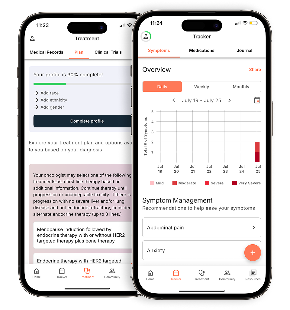
| Feature | Type 1 | Type 2 |
|---|---|---|
| Cell size | Small | Large |
| Cytoplasm | Scant, pale | Abundant, eosinophilic (pink) |
| Chromosome alterations | Gains of chromosomes 7 and 17, loss of Y | Gains of chromosome 1q, complex alterations |
| Key gene association | MET proto-oncogene | FH (fumarate hydratase) gene mutations are associated with a hereditary form of renal cell carcinoma that can present with papillary features, though the specific connection to Type 2 papillary RCC classification has not been clearly established |
MET proto-oncogene mutations, both hereditary and sporadic, are closely linked to papillary RCC Type 1, driving abnormal cell proliferation through the MET signaling pathway. These molecular differences directly shape which targeted therapies your care team may consider. Understanding how these subtypes behave at the cellular level sets the stage for examining how differently they grow and spread.
Evaluating growth patterns and aggression
Type 1 papillary renal cell (kidney) cancer tends to grow slowly and remain localized, while Type 2 behaves far more aggressively, often spreading before it’s detected. This distinction directly shapes papillary renal cell carcinoma treatment decisions and urgency of intervention. Type 1 papillary renal cell carcinoma tumors are typically indolent, meaning your care team may have more time to monitor and plan. Type 2, by contrast, tends to metastasize early, most commonly to the lymph nodes and lungs, making prompt action critical. These contrasting behaviors are a key reason subtype identification matters so much at diagnosis, a point that becomes even clearer when you look at survival outcomes across subtypes.
Which renal cell carcinoma has the worst prognosis?
Subtype and survival. Research comparing outcomes across both types shows meaningful differences. Type 1 papillary renal cell carcinoma caught at an early stage generally shows high rates of progression-free and cancer-specific survival, though specific survival percentages vary depending on other tumor features. Type 2 papillary renal cell carcinoma has a median overall survival of approximately 12.6 months in patients with metastatic disease, which is shorter than the 20.0 months observed in type 1 papillary renal cell carcinoma, though this difference was not statistically significant in the available data. Type 2 pRCC often presents at an advanced stage. A study confirming that Type 2 histology independently predicts worse oncological outcomes across multiple metrics underscores this pattern.
Stage at diagnosis. Subtype alone doesn’t determine risk; stage at diagnosis amplifies it. A patient with Type 2 pRCC identified early may still benefit from curative surgery. But because Type 2 tends to spread before symptoms appear, localized detection is far less common, and that interaction between subtype and stage compounds the prognosis significantly.
Recurrence risk. Type 2 papillary renal cell carcinoma may be associated with a higher risk of cancer recurrence compared to type 1, and your care team can discuss what this means for your individual outlook and follow-up plan. Even after successful resection, the probability of the disease returning remains elevated compared to Type 1. This reinforces why your care team’s monitoring strategy after treatment matters as much as the initial intervention. Those differences in long-term outcomes are precisely what the next section examines in detail.
Survival rate for papillary renal cell (kidney) cancer Type 2
Fuhrman grade. Fuhrman grade plays a central role in survival differences between Type 1 and Type 2 papillary renal cell carcinoma. Type 2 papillary renal cell carcinoma has been considered more aggressive, but recent research suggests that the presence or amount of type 2 features does not significantly predict patient outcomes differently from type 1 tumors. That cellular aggression translates into faster progression and a reduced response to treatment.
Early detection remains the most meaningful lever for improving outcomes. When Type 2 is caught before it spreads beyond the kidney, surgical intervention can still be effective, making routine imaging and prompt evaluation critical. Subtype goes only so far in predicting prognosis, though. Factors like tumor size, staging, and surgical margins also shape the overall picture, which the next section examines in detail.
Prognostic indicators beyond subtyping
Subtype alone doesn’t determine outlook; tumor size, staging, and pathological features combine to shape the full prognostic picture for renal cell (kidney) cancer.
Cancer care guidance for every step of your journey
Get treatment options, clinical trials, and support tailored to your diagnosis--all in one place.
Get started
See treatment options, manage symptoms, and stay informed—all in the app
View treatment options and trials personalized to your diagnosis—plus track progress in real time.
Continue in appTNM staging and tumor size remain foundational. Localized disease caught early carries far better odds than metastatic presentation, regardless of subtype. But in Type 2, sarcomatoid differentiation, where cancer cells take on an aggressive, spindle-shaped form, can accelerate disease progression significantly, compounding an already guarded prognosis. Surgical margins and lymphovascular invasion also matter: lymphovascular invasion at the time of surgery in papillary renal cell carcinoma is associated with reduced cancer-specific survival.
Understanding these layered indicators sets the stage for a closer look at how treatment decisions, from surgery type to systemic therapy, must adapt to each patient’s specific profile.
Navigating treatment for kidney cancer subtypes
Treatment for renal cell (kidney) cancer is shaped by the specific subtype, its genetic profile, and how far it’s spread, all of which inform a personalized care plan.
Surgical approach remains the primary treatment for localized papillary renal cell (kidney) cancer, but the choice between partial and radical nephrectomy depends heavily on subtype aggression. Type 1 papillary renal cell (kidney) cancer tumors tend to be slow-growing, which may make them suitable candidates for certain treatment approaches including surgery. Type 2 papillary renal cell carcinoma tumors tend to be more aggressive than type 1 tumors, which may affect treatment decisions your doctor makes.
Systemic therapies diverge sharply between subtypes once disease extends beyond the kidney. The WHO 2022 classification recognized that type 2 papillary renal cell cancer includes entities with different molecular backgrounds that may overlap with other renal tumor types. Targeted agents and immunotherapy combinations are being studied as treatment options for advanced papillary renal cell cancer, including in clinical trials combining agents like nivolumab and cabozantinib. Type 1 papillary renal cell carcinoma tumors frequently harbor MET pathway alterations, making MET-targeted therapies an area of active clinical investigation.
Genetic profiling is increasingly central to building a treatment strategy. Rather than applying a broad treatment framework, care teams now use tumor genomics to identify actionable mutations, a shift that has moved papillary renal cell (kidney) cancer management firmly toward individualized decision-making. How that plays out in practice often starts at the surgical planning stage, which is where questions around active surveillance and operative approach become critically important.
Surgical standards and active surveillance
Surgical decisions for papillary renal cell (kidney) cancer hinge on subtype, tumor size, and the risk profile your care team has established through staging and pathological review.
Active surveillance is a viable path for small, incidentally discovered Type 1 tumors, particularly those under three centimeters with low-grade features. Type 1 papillary renal cell carcinoma tends to grow slowly, so a watchful waiting approach can spare you unnecessary intervention while your care team monitors for any meaningful progression.
Type 2 lesions call for a more aggressive response. Wide surgical margins are prioritized to reduce the risk of local recurrence. Type 2 papillary renal cell carcinoma tends to have more aggressive features than Type 1, which is why your surgical team may discuss whether wider surgical margins could be part of your treatment plan.
Robotic-assisted vs. open surgery is a practical consideration for complex pRCC cases. Robotic-assisted techniques offer precision and faster recovery for many patients, but open surgery remains the standard when tumor size, location, or vascular involvement makes minimally invasive approaches less reliable. Your care team will weigh both options carefully against your personalized care plan. When systemic therapy enters the picture, which it often does for advanced or high-risk cases, the choice of surgical approach becomes just one part of a broader treatment strategy, leading into the critical role of immunotherapy and targeted agents.
Systemic therapies. Immunotherapy and targeted agents
Subtype classification directly shapes which systemic therapies your care team will consider, and why agents that work well for clear cell renal cell (kidney) cancer often fall short for papillary disease.
MET pathway alterations are common in papillary renal cell carcinoma, occurring in all familial cases and approximately 13% of sporadic cases, which may have implications for treatment selection. Type 2 papillary renal cell carcinoma tends to have a more aggressive profile and higher immune activity than Type 1, which may make checkpoint inhibitors targeting PD-1/PD-L1 worth discussing with your care team as a possible treatment option. Standard agents like sunitinib, developed primarily around clear cell biology, tend to produce weaker responses in papillary subtypes, which is why subtype-specific personalized care plans matter so much in advanced disease.
That said, the molecular picture within papillary renal cell (kidney) cancer is more complex than subtype labels alone can capture, a nuance the next section explores in depth.
Limitations and considerations in pRCC classification
The Type 1 vs. Type 2 framework is clinically useful, but it was never designed to capture every tumor, and its limitations are becoming harder to ignore as molecular testing reshapes how your care team understands renal cell (kidney) cancer.
Mixed and unclassified features complicate diagnosis more often than the clean Type 1/Type 2 split suggests. Some papillary renal cell carcinoma tumors display overlapping papillary architecture that doesn’t fit neatly into either Type 1 or Type 2 category, landing in an unclassified RCC designation, leaving both pathologists and clinicians working with incomplete information.
Needle core biopsies add another layer of uncertainty. Needle core biopsies of renal tumors may not capture all of the diagnostic and prognostic information needed to fully characterize the tumor, and additional molecular and genetic analysis of biopsy specimens can provide information to support treatment decisions. This matters enormously when different regions of the same papillary renal cell carcinoma tumor can behave differently. A biopsy that misses a more aggressive area can lead to an incomplete picture of what you’re actually dealing with, a problem the next section explores in depth.
The 2022 WHO classification reflects how far the field has moved. It de-emphasizes the simple Type 1/Type 2 binary in favor of molecularly defined subtypes, recognizing that visual morphology alone doesn’t reliably predict behavior or treatment response. This shift signals a broader transition. Molecular profiling is becoming as important as, and in some cases more important than, what a tumor looks like under a microscope.
The challenge of intratumoral heterogeneity
A single renal cell (kidney) cancer tumor can harbor both Type 1 and Type 2 regions simultaneously, meaning a biopsy sample may not reflect the full biological picture. This phenomenon, called intratumoral heterogeneity, creates a real diagnostic risk. If sampling misses the more aggressive Type 2 component, the tumor may be under-graded. Your care team would then be working from an incomplete picture when building your personalized care plan. Because of this, clinicians typically treat papillary renal cell carcinoma based on the most aggressive feature identified, not the average.
The shift to molecular pathology
Histologic subtyping alone can’t always predict how a renal cell (kidney) cancer will behave, and that gap is driving a critical shift toward molecular pathology.
Some Type 2 papillary renal cell carcinoma tumors grow slowly despite their aggressive appearance, while some Type 1 papillary renal cell carcinoma cases progress faster than expected. When clinical behavior doesn’t match the morphology, looking at underlying metabolic pathways, such as MET signaling or fumarate hydratase deficiency, provides a clearer picture. If your pathology results feel inconclusive or your treatment response is unexpected, asking your care team for a referral to a specialized urologic pathologist is a reasonable and informed next step. With that fuller picture in hand, the key takeaways ahead can help you frame the right questions.
Key takeaways: what you need to know
Understanding the difference between Type 1 and Type 2 papillary renal cell (kidney) cancer directly shapes your prognosis, your treatment options, and the decisions you make with your care team.
Here’s what to carry forward from this article:
- Type 1 pRCC tends to be slow-growing with a more favorable long-term survival rate, making active surveillance a realistic option in select cases.
- Type 2 pRCC tends to be more aggressive than type 1, and its behavior can be harder to predict.
- Molecular and genetic testing has become critical for accurate subtyping, histology alone doesn’t always tell the full story, and your personalized care plan should reflect that precision.
- Your pathology results matter. Discuss them in detail with your care team so every treatment decision is grounded in your specific biology.
Disclaimer: The information provided in this article is for informational purposes only and is not intended to be a substitute for professional medical advice, diagnosis, or treatment. Outcomes4Me is not acting as your caregiver, and any suggestions or guidance offered should not replace the advice of your healthcare provider or qualified medical professional. Always seek the guidance of your physician or other qualified health provider with any questions you may have regarding a medical condition.
Personalized support for real care decisions
Understand your diagnosis, explore clinical trials, and track symptoms--all in one place.
Get started
Compare treatments, prepare for appointments, and track side effects—all in the app
Built for your diagnosis, Outcomes4Me gives you the tools to make confident, informed decisions—right when you need them.
Continue in app