Funding for the Lung Health Hub is provided by AstraZeneca. All content is developed independently by Outcomes4Me; AstraZeneca has no influence on the content of the site.
If you’ve been told you have a lung nodule, your care team will guide you through next steps tailored to the characteristics of the nodule and your personal risk factors. Your care team will conduct additional follow-up tests to provide your doctor with more information and clarity. In this section, we’ll cover the different ways your care team will evaluate lung nodules and the additional imaging you may need.
Personalizing the evaluation process
Not all lung nodules are the same. Below is a quick recap of what your care team will consider when deciding the best approach for you:
- Nodule size: Smaller nodules are less likely to be cancerous and often only need periodic monitoring with repeat scans. Larger nodules may require further evaluation.
- Nodule density: Nodules can be solid or subsolid. Solid nodules appear dense on scans, while subsolid nodules have a hazy or fuzzy appearance.
- Nodule shape: Nodules can be round, smooth, bumpy, or spikey.
- Risk factors: Your medical history and risk profile play a big role. Factors such as smoking history, age, family history of lung cancer, or previous personal cancer diagnosis influence the level of concern and the recommended next steps.
What happens after you find out you have a lung nodule from a CT scan or X-ray?
Based on these factors, your doctor may recommend:
Watchful waiting
For small, low-risk nodules,your doctor may schedule regular CT (computed tomography) scans to monitor any changes in size or appearance over time. During the scan, you’ll lie on a narrow table that slides in and out of a machine. You may be asked to hold your breath for a few seconds to help get clear images. The process is quick, painless, and typically takes less than 10 minutes. Depending on your nodule characteristics, these scans are repeated anywhere from every 3-6 months for two years.
Further imaging
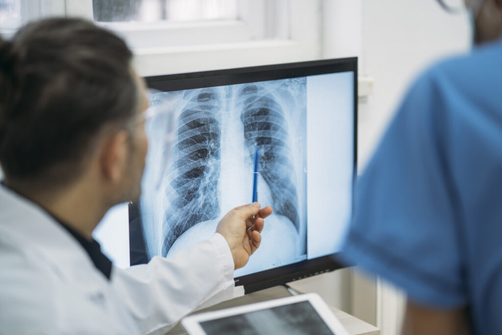
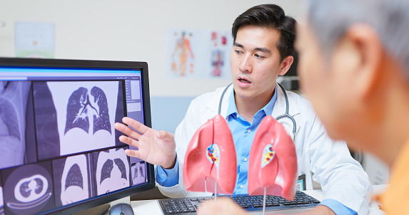
A PET/CT (positron emission tomography/computed tomography) scan can evaluate nodules that look suspicious on CT or have grown. This scan helps determine if the nodule is metabolically active (which can indicate cancer) by showing areas of increased glucose uptake. Before the scan, you’ll receive a small injection of a radioactive sugar solution. Then, you’ll lie down on a table where the PET/CT scanner will take detailed images that show both the structure of your lungs and areas of increased metabolic activity. The scan itself usually takes about 20-30 minutes.
Biopsy for tissue sample
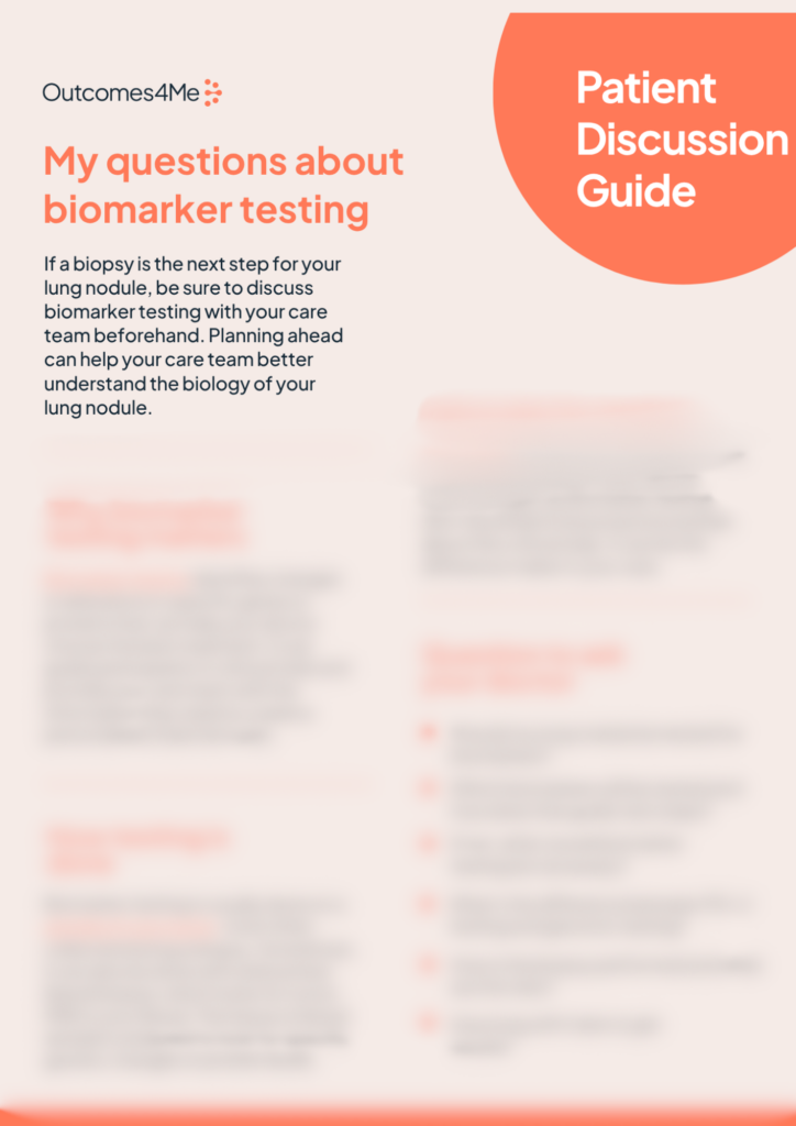
If the nodule appears suspicious for cancer, your doctor may suggest a biopsy to obtain a tissue sample for diagnosis. There are several ways this can be done, depending on the size and location of the nodule and your overall health. If you’d like more information on the different procedures of tissue removal, download our Patient Decision Aid: “Is a biopsy the next step?” which goes over the methods available.
Referral to specialists
Depending on the findings, you may receive a referral to a pulmonologist, thoracic surgeon, or oncologist to discuss diagnosis and treatment options. If you have questions or concerns about your lung nodule or the follow-up plan, don’t hesitate to reach out to your healthcare providers.
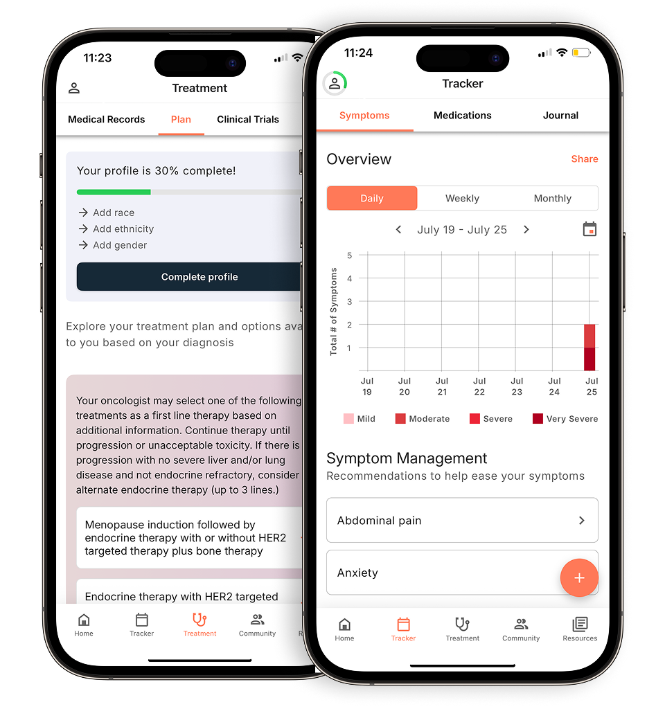
Personalized support for real care decisions
Understand your diagnosis, explore clinical trials, and track symptoms--all in one place.
Get started
Compare treatments, prepare for appointments, and track side effects—all in the app
Built for your diagnosis, Outcomes4Me gives you the tools to make confident, informed decisions—right when you need them.
Continue in app