The 2024 FDA mandate: why your mammogram letter just changed
If you received a mammogram recently and noticed something different in your results letter, you’re not imagining it. As of September 2024, every mammography facility in the U.S. is legally required to tell you whether you have dense breast tissue — in plain, standardized language you can actually understand.
This change stems from the FDA’s update to the Mammography Quality Standards Act (MQSA), which took full effect in September 2024. Before this mandate, whether you learned about your breast density depended largely on where you lived. Some states had already enacted their own notification laws, but many hadn’t, creating a geographic lottery that left millions of patients without information that directly affects how their screening results should be interpreted.
The new rule addresses this inconsistency. Under the updated dense breast tissue mammogram guidelines, facilities must now use federally standardized language in every patient summary. If your tissue is dense, your letter will say so explicitly, and it will note that dense tissue can make it harder to detect cancer on a mammogram alone.
It’s crucial to understand the scope of this mandate. It ensures you receive the information. What happens next — whether you pursue additional screening options or discuss your personal risk profile with your care team — still requires a conversation. Think of the notification as the starting point, not the full picture. To fully act on it, you first need to understand what breast density actually means, and that starts with knowing which category applies to you.
Decoding the four categories of breast density
Breast density is not a sensation, it’s a radiologic finding that only shows up on an imaging report. You can’t determine your density by how firm or soft your breasts feel during a self-exam or a clinical check. What radiologists are actually measuring is the ratio of fibroglandular tissue to fatty tissue visible on a mammogram. Breast density is classified into four categories that range from almost all fatty tissue to extremely dense tissue with very little fat, helping radiologists communicate findings on mammogram reports.
Understanding where you fall on the BI-RADS scale matters for interpreting your results letter and understanding what your radiologist found on your breast imaging exam.
- Category A – Entirely fatty: The breast is composed almost entirely of fat. Mammograms are highly sensitive in this tissue type, and masses are relatively easy to spot.
- Category B – Scattered fibroglandular density: Some fibroglandular tissue is present, but fat remains the dominant tissue. This is one of the most common findings and carries low concern.
- Category C – Heterogeneously dense: Fibroglandular tissue is distributed unevenly throughout the breast and may obscure small masses, making interpretation more complex.
- Category D – Extremely dense: The breast contains a very high proportion of fibroglandular tissue relative to fat. This significantly reduces mammogram sensitivity.
Categories C and D are the threshold that triggers the FDA’s new notification requirement — together, they account for nearly half of all women who get mammograms. If your letter mentions “heterogeneously dense” or “extremely dense” tissue, you’re in this group. That distinction is clinically meaningful, and understanding why requires a closer look at exactly what happens to a mammogram image when dense tissue is involved.
Evidence-based guidance powered by NCCN Guidelines®
Personalized treatment plans shaped by the latest oncology standards—tailored to your diagnosis.
Get started
View your personalized treatment plan in the Outcomes4Me app
Use your diagnosis to unlock personalized NCCN Guidelines®-aligned recommendations.
Continue in app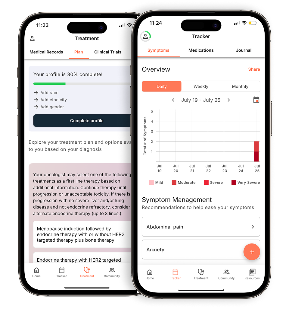
The masking effect: why mammograms alone aren’t enough
Dense tissue not only raises your cancer risk, it actively hides the evidence of it, and that distinction matters enormously for how you approach screening.
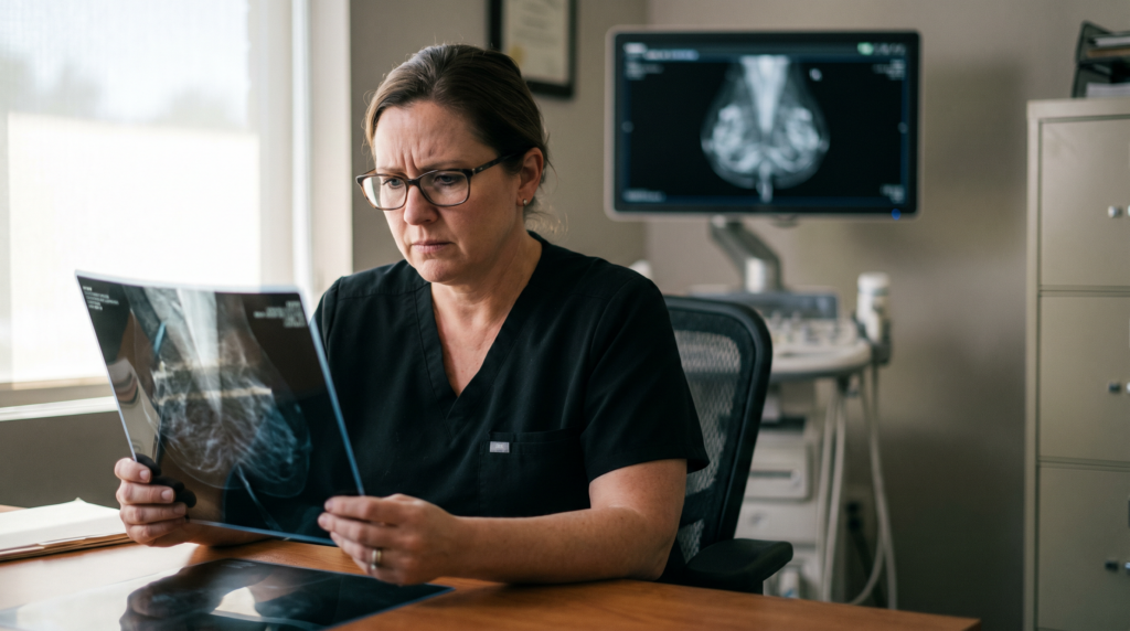
On a standard mammogram, X-rays pass through the breast and produce a grayscale image. Fatty tissue appears dark, which is why tumors, which appear white, stand out clearly against it. But fibroglandular tissue also appears white. In a dense breast, a tumor and the surrounding tissue can be virtually indistinguishable, like trying to spot a white marble on a white tablecloth. Radiologists call this the masking effect, and it’s the primary reason cancers are missed in women with heterogeneously or extremely dense breasts.
“The challenge with dense breast tissue is not due to mammogram quality, it’s a physics problem. You’re looking for a white finding in a white background, and that’s genuinely difficult even for experienced radiologists.” — perspective shared broadly across breast imaging specialists
The stakes here go beyond a technical imaging limitation. Dense breast tissue can make it slightly more difficult for mammograms to detect cancer, though modern imaging techniques like 3D mammography remain effective screening tools even in women with dense breasts. Meanwhile, women with extremely dense breasts face an elevated risk of developing breast cancer compared to those with predominantly fatty tissue, according to the Breast Cancer Research Foundation. That’s a compounding problem: the patients most at risk are also the ones for whom the standard screening tool performs least reliably.
This is precisely why the FDA breast density notification requirement exists — not to alarm patients, but to make sure they understand that a “normal” result on a mammogram isn’t always the complete picture. A Category D patient who receives a clear mammogram report hasn’t necessarily been given an all-clear. She may simply have received an incomplete one. Understanding how different imaging tools work is a critical next step, because for many women with dense tissue, supplemental screening isn’t optional, it’s essential.
Navigating supplemental screening: ultrasound vs. MRI
Understanding your breast density category is just the beginning, the harder question is what you should actually do with that information when it comes to supplemental screening.
3D mammography (tomosynthesis) has become the new standard, but it still has real limits in dense tissue. It captures multiple image slices rather than a single flat image, which improves detection compared to traditional 2D mammography. However, heterogeneously or extremely dense tissue can still obscure small tumors, which is exactly why the breast density categories C and D in your report often prompt a conversation about going further.
Cancer care guidance for every step of your journey
Get treatment options, clinical trials, and support tailored to your diagnosis--all in one place.
Get started
See treatment options, manage symptoms, and stay informed—all in the app
View treatment options and trials personalized to your diagnosis—plus track progress in real time.
Continue in appWhen supplemental screening is recommended for dense breast tissue, options may include breast ultrasound or MRI, which can be discussed with your healthcare provider based on your individual risk factors and health history:
- Handheld or automated breast ultrasound (ABUS) is typically the first supplemental step for women with dense tissue and average-to-intermediate risk. It’s widely available, doesn’t use radiation, and can detect cancers missed on mammography, though it does carry a higher rate of false positives.
- Breast MRI is considered the gold standard for high-risk patients, generally those with a lifetime risk above 20%, but it requires specific clinical indications to access and is not routinely recommended for all women with dense breasts alone.
- Contrast-enhanced mammography (CEM) is an emerging middle-ground option for patients who can’t tolerate MRI, offering improved sensitivity over standard mammography with lower cost and broader availability.
It’s worth noting that the USPSTF 2024 Guidelines issued an “I Statement” on supplemental screening, acknowledging that while these additional tools do find more cancers, the evidence on long-term outcomes is still developing. That’s not a reason to avoid supplemental screening; it’s a reason to make decisions grounded in your full risk picture, not density alone.
That full picture, including family history, genetics, and lifetime risk scores, is what the next section addresses.
Moving toward personalized risk assessment
Breast density is a critical screening variable, but it’s only one piece of a much larger picture, and treating it in isolation can give you an incomplete view of your actual risk.
Family history, genetic markers like BRCA1 and BRCA2, age at first menstrual period, and prior biopsy results all interact with density to shape your individual cancer profile. This is why the conversation with your provider shouldn’t stop at density category. Asking specifically for a formal risk model, such as Tyrer-Cuzick or the Gail Model, is one of the most actionable steps you can take. According to evidence-based guidelines, validated risk-assessment tools are available that incorporate patient-specific data, including age, family history, and breast tissue density, to help guide personalized breast cancer screening and management strategies.
Here’s why: most insurance providers, including Medicare, require a documented lifetime risk of 20% or higher before they’ll cover breast MRI. Without a formal risk assessment on file, you may face out-of-pocket costs even when supplemental screening is clinically appropriate. Similarly, the ongoing debate around imaging technology, including whether a 3D mammogram vs 2D for dense breasts offers meaningfully better detection, often hinges on individual risk stratification, not blanket recommendations.
Pulling all of this together, your density category, genetic history, screening results, and risk score, is where many patients get stuck. Understanding your breast density and screening results is an important part of managing your breast health, particularly as you navigate follow-up appointments and additional imaging recommendations. Understanding your full picture is the foundation for the practical steps covered in the next section.
The bottom line: your action plan for dense breasts
Notification is the first step, but as the Outcomes4Me Editorial Board notes, “personalized advocacy is what saves lives in the context of dense breast tissue.” The new mammogram laws 2024 put standardized density reporting in place nationwide, yet that information only becomes protective when you act on it.
Here’s a clear framework for what to do next:
- Check your BI-RADS category. Your mammogram report will list one of four density categories (A through D). Review your breast cancer screening options with this category in hand, it anchors every conversation that follows.
- Schedule a targeted follow-up if you’re Category C or D. Don’t wait for your next annual appointment. Ask specifically about supplemental screening options, such as ultrasound or MRI, based on your density level.
- Request a formal risk assessment. Risk assessment tools like the Tyrer-Cuzick model can help identify women who may benefit from supplemental screening beyond mammography, particularly those with dense breasts.
- Use digital health tools to stay organized. Tracking your screening history, results, and upcoming appointments in one place ensures nothing falls through the cracks, and keeps you current on clinical trials evaluating next-generation imaging technologies.
Dense breast tissue requires a proactive, structured response, not just a letter in the mail.
The next section offers specific questions to bring to your provider so you can walk into that appointment prepared and informed.
Advocating for your care: questions for your next appointment
Knowing your density category is only valuable if you act on it, and that starts with asking the right questions at your next appointment. The patients who get the most out of their screening are those who walk in prepared, not those who wait to be told what they need.
Start with these five questions:
- “Given my density category, what is the likelihood a mammogram alone would miss a finding?”
- “What is my lifetime risk score, and does it qualify me for supplemental MRI?”
- “Does my current insurance plan cover automated breast ultrasound (ABUS) or other supplemental screening?”
- “Should I be screened annually, or does my risk profile suggest a different schedule?” (For context on how screening intervals are being debated, the 2024 guidelines landscape is still evolving.)
- “Are there clinical trials studying supplemental screening approaches that I might be eligible for?”
Empowerment comes from organizing your data and being prepared before your appointment. Proposed federal legislation, the Find It Early Act, aims to remove cost-sharing barriers for supplemental screening entirely, but until that passes, knowing exactly what your plan covers puts you in a stronger negotiating position.
Outcomes4Me helps you bring all of this together. You can discuss your breast density screening results and access resources about supplemental screening options, including information about ultrasound and MRI, to help you make informed decisions about your care with your medical team. The 2024 FDA mandate gave you the notification. Outcomes4Me helps you figure out what to do next.
Personalized support for real care decisions
Understand your diagnosis, explore clinical trials, and track symptoms--all in one place.
Get started
Compare treatments, prepare for appointments, and track side effects—all in the app
Built for your diagnosis, Outcomes4Me gives you the tools to make confident, informed decisions—right when you need them.
Continue in app