Recognizing the unique presentation of inflammatory breast cancer (IBC)
IBC is an aggressive form of breast cancer that is thought to involve cancer cells blocking lymph vessels in the skin of the breast, which may contribute to the characteristic skin changes. Because the tumor grows in sheets and nests rather than a single consolidated mass, there’s often nothing firm to find during a self-exam. The disease announces itself through the skin instead.
Timing is the other distinguishing feature. Where many breast cancer symptoms develop slowly over months, IBC skin changes may develop relatively rapidly, with some patients noticing changes over days to weeks, though the timeline can vary. Rapid onset combined with warmth and redness is precisely why IBC is mistaken for an infection, and why symptoms that look like a simple breast infection deserve prompt, documented evaluation rather than watchful waiting.
Visual hallmarks. Peau d’orange and color shifts
One hallmark appearance of inflammatory breast cancer is a skin texture sometimes called peau d’orange, which may resemble an orange rind. This dimpled or pitted appearance is thought to result from swelling related to lymphatic changes, though the exact appearance can vary from person to person. Running a hand across it, peau d’orange breast feels thickened and slightly firm rather than soft.
Color matters, and it varies. On lighter skin, breast skin discoloration in IBC may appear as bright pink or red, though the specific appearance can vary from person to person. On deeper skin tones, the inflammatory changes may appear purple, dusky brown, or bruised-looking, though the appearance can vary from person to person. IBC redness is usually diffuse and patchy across a broad area, unlike the tight, well-defined border of a localized rash.
Nipple changes may occur with IBC and can include sudden flattening, inversion, retraction, or crusting, often on one side only. If you notice these changes, it’s important to mention them to your doctor.
IBC vs. mastitis: comparing early symptoms
The two conditions overlap enough that a careful comparison is the most useful diagnostic tool a patient has.
Evidence-based guidance powered by NCCN Guidelines®
Personalized treatment plans shaped by the latest oncology standards—tailored to your diagnosis.
Get started
View your personalized treatment plan in the Outcomes4Me app
Use your diagnosis to unlock personalized NCCN Guidelines®-aligned recommendations.
Continue in app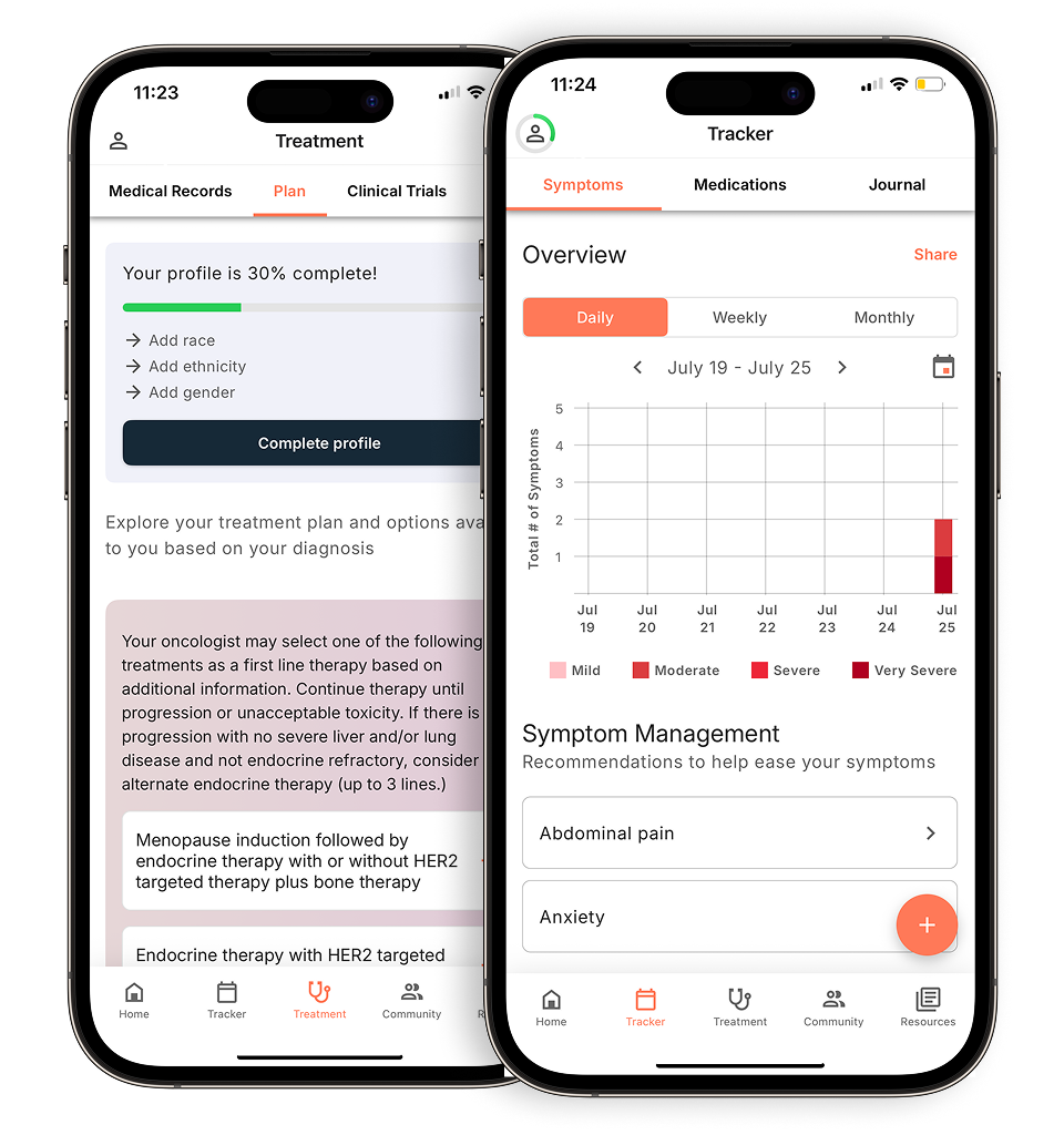
| Feature | Mastitis | Inflammatory breast cancer |
|---|---|---|
| Onset | May develop suddenly, sometimes in connection with lactation or a blocked duct, though inflammatory breast cancer can occur in other circumstances as well | Typically develops suddenly. While it can occur in people who are nursing, it is not caused by nursing itself |
| Fever and chills | Fever and chills may occur with mastitis and can sometimes be part of a broader illness response, though they are not always present in every case | Fever and chills may not be present in the early stages of IBC, though symptoms can vary from person to person |
| Skin texture | Some people with mastitis describe the skin as feeling tender and warm, and it may appear shiny | IBC skin may appear thickened, pitted, or ridged, sometimes described by patients as having a heavy or dimpled texture similar to an orange peel |
| Response to antibiotics | Mastitis often shows improvement within days when treated with antibiotics, though response times can vary from patient to patient | IBC may not respond to antibiotics the way typical skin infections do, which is one reason prompt evaluation by a doctor is important if you notice these symptoms |
| Pattern | In inflammatory breast cancer, skin changes may appear in a wedge or quadrant pattern, though the appearance can vary from person to person | IBC redness and swelling often appear across a larger area of the breast rather than in one spot, and may continue to spread over time |
Skin changes from infection may improve with antibiotics, whereas those from malignant lymphatic obstruction typically do not. This difference can sometimes help your care team distinguish between the two conditions. If a course finishes and the breast redness and swelling is unchanged or worse, that non-response is clinical information, not a reason for a second course.
Feel is another clue. An inflamed breast from infection may feel acutely painful and warm or hot to the touch, though these sensations can vary from person to person. IBC may produce a sense of density, weight, and stretched skin. Some patients report breast itching and burning alongside or instead of sharp pain.
Diagnostic pathways for skin-only symptoms
A normal mammogram does not necessarily rule out IBC, so it’s important to mention any skin changes to your doctor even if imaging results appear normal. Diffuse skin and lymphatic disease may produce no discrete mass to image, and an edematous, tender breast can be difficult to compress adequately, so the study can read as unremarkable while disease is present. Treating that result as an all-clear is one of the most consequential errors in this diagnosis.
Ultrasound is better suited to the question. Ultrasound may be used to look for skin thickening, dilated dermal lymphatics, architectural distortion, and enlarged axillary nodes, which can sometimes be seen in IBC. Your care team can explain whether ultrasound might be helpful in your specific situation. Breast MRI may be used as part of the imaging workup for IBC to help evaluate the extent of involvement in breast tissue and lymph nodes. Your care team can discuss whether this imaging is appropriate for your situation.
Cancer care guidance for every step of your journey
Get treatment options, clinical trials, and support tailored to your diagnosis--all in one place.
Get started
See treatment options, manage symptoms, and stay informed—all in the app
View treatment options and trials personalized to your diagnosis—plus track progress in real time.
Continue in appA skin punch biopsy of the affected area is often used as part of the diagnostic process for IBC, as it can help confirm the diagnosis. Core biopsy of any suspicious parenchymal or nodal finding runs in parallel, along with receptor testing. Your care team may recommend receptor testing (hormone receptor and HER2 status) to help inform treatment options. Ask your doctor whether this testing is appropriate for your diagnosis. Doctors typically look at how IBC presents and how quickly it develops when considering a diagnosis, and a biopsy may be used to help confirm the findings.
Common Questions About IBC Progression
How long can you have IBC without knowing? IBC may progress rapidly, with some patients experiencing noticeable changes over a period of weeks rather than months. The progression window is compressed, which is both the danger and the diagnostic clue. Anything that transforms a breast’s appearance in under a month warrants urgent assessment.
What is mistaken for inflammatory breast cancer? IBC can be mistaken for other conditions affecting the breast and skin, such as mastitis, breast abscess, cellulitis, eczema, contact dermatitis, radiation dermatitis, and insect reactions. If you have persistent skin changes on your breast, it’s important to see a doctor for proper evaluation, as these conditions require different treatments.
Why is IBC diagnosed relatively late? Two reasons compound each other. There’s usually no lump to trigger the familiar screening reflex, and the visible signs of breast cancer here mimic benign conditions convincingly enough that empiric treatment feels reasonable. Time spent confirming a benign explanation is time the disease uses.
Limitations and considerations in identification
Visual presentation varies considerably between individuals. Some breasts show florid peau d’orange; others show only subtle asymmetric firmness and faint discoloration. The amount of redness visible on the skin may not always match how far the disease has progressed, so it’s important not to assume severity based on appearance alone. Not all cases of IBC show the classic orange-peel texture, so this skin change alone shouldn’t be used to rule IBC in or out. If you notice any skin changes on your breast, talk with your doctor about what you’re seeing.
Online galleries have real limits. Searching for pictures of inflammatory breast cancer in early stages or early signs of breast cancer pictures can help you name what you’re seeing and describe it precisely to your care team. What those images can’t do is confirm or exclude a diagnosis, because photographs flatten texture, distort color under varied lighting, and underrepresent deeper skin tones. Use them as a reference, not as a diagnosis.
Prior breast surgery adds another layer. Scarring, implants, reduction, or reconstruction can alter skin contour, contract tissue, and produce baseline thickening that masks or mimics early change. Comparing to your own prior photographs is more informative than comparing to someone else’s.
One final caution. Some people with IBC may not experience pain, even though skin changes are present, though pain experiences vary widely among patients. Comfort is not evidence of a benign process, and the absence of pain should not delay evaluation.
When to consult a medical provider
If a breast rash or presumed infection hasn’t clearly improved after roughly seven days of appropriate treatment, return for reassessment and ask directly about IBC.
If imaging is inconclusive but symptoms continue, request a skin punch biopsy by name and ask that IBC be documented as part of the differential in your chart. Written questions make that conversation easier.
Report these findings specifically:
- Pitted or dimpled skin texture
- Sudden one-sided swelling or size increase
- A breast that feels hot to touch, persistently
- Ridged or thickened skin
- New nipple flattening, inversion, or crusting
What you need to know
- IBC presents through the skin, redness, pitting, thickening, and swelling, rather than through a palpable lump, so a negative self-exam means little here.
- Early symptoms of breast cancer in this form are routinely misidentified as mastitis, cellulitis, or dermatitis; failure to respond to antibiotics is meaningful.
- A skin punch biopsy showing tumor cells in the dermal lymphatics is the definitive confirmation, and a normal mammogram doesn’t exclude the diagnosis.
- Because progression is measured in weeks, prompt evaluation changes what treatment options remain available.
The Outcomes4Me app references National Comprehensive Cancer Network® (NCCN®) treatment guidelines, which may help you organize information about treatment options to discuss with your care team.
Disclaimer: The information provided in this article is for informational purposes only and is not intended to be a substitute for professional medical advice, diagnosis, or treatment. Outcomes4Me is not acting as your caregiver, and any suggestions or guidance offered should not replace the advice of your healthcare provider or qualified medical professional. Always seek the guidance of your physician or other qualified health provider with any questions you may have regarding a medical condition.
Personalized support for real care decisions
Understand your diagnosis, explore clinical trials, and track symptoms--all in one place.
Get started
Compare treatments, prepare for appointments, and track side effects—all in the app
Built for your diagnosis, Outcomes4Me gives you the tools to make confident, informed decisions—right when you need them.
Continue in app