
A CA-125 result below 35 U/mL doesn’t rule out ovarian cancer, which is one of the most disorienting things patients face after a diagnosis.
CA-125 (cancer antigen 125) is a protein shed by certain cells, including some ovarian cancer cells, that circulates in the bloodstream. Laboratories use a threshold of less than 35 U/mL as the standard “normal” range, but that number is a statistical baseline, not a diagnostic tool. It reflects what’s typical in a healthy population, not a guarantee that cancer is absent.
This is where the emotional challenges run deepest. Research shows that up to 20% of advanced ovarian cancers and roughly 50% of early-stage cases produce normal CA-125 levels. That gap between a number and a reality is genuinely hard to process.
It’s also important to understand what this biomarker is and isn’t. CA-125 is most valuable as a monitoring tool, tracking how your cancer responds to treatment or detecting recurrence over time. CA-125 was never validated as a standalone screening tool for the general population. Your care team will interpret it alongside imaging, pathology, and your full clinical picture. Understanding the standard reference range in more detail is the essential next step.
Understanding the standard reference range
Many US laboratories use a threshold of 0–35 U/mL to define a normal CA-125 range, though this cutoff can vary between laboratories and may not apply equally to all populations. Labs set this range using large reference studies, though values can shift slightly depending on the specific assay a facility uses and a patient’s age. What matters more than any single reading is your baseline, a consistent pattern of results tracked over time. A CA-125 of 30 U/mL rising steadily across multiple tests can signal more concern than an isolated result of 40 U/mL. Your provider will order CA-125 tests in a series so they can identify changes in your levels over time, rather than relying on a single test result. And understanding that distinction sets the stage for a closer look at just how often “normal” can mislead.
The diagnostic limitations of CA-125
CA-125 alone detects only about 62% of early-stage ovarian cancers at high specificity, meaning it misses roughly 38% of cases in this stage. This limitation shapes ovarian cancer prognosis from the very first appointment.
Tumor subtype plays a significant role here. Some ovarian cancers, including mucinous tumors, can have normal CA-125 levels even when the disease is advanced, meaning the bloodstream shows nothing unusual even when disease is present. And in premenopausal women, endometriosis and menstruation can cause elevated CA-125 levels, driving a high rate of false positives that complicates interpretation further. That raises an important question. If normal results don’t confirm absence of disease, what does drive a tumor to stay silent on this test?
Why some ovarian cancers don’t trigger a high CA-125
Some ovarian tumors don’t shed the CA-125 antigen into the bloodstream in significant amounts, which helps explain how ovarian cancer diagnosis can be affected by a delayed or missed diagnosis.
Non-secretor tumors are the primary reason a cancer can be active while the CA-125 test returns a completely normal result. Some tumor cells simply don’t express the gene that produces the CA-125 protein at detectable levels, meaning the antigen never reaches the bloodstream in significant quantities. This is a biological characteristic of the tumor itself, not a reflection of disease severity.
Tumor stage and FIGO grading also shape how much protein a cancer produces. Early-stage tumors, particularly FIGO Stage 1, are often confined to one or both ovaries with minimal surface involvement, so they produce far less CA-125 than advanced-stage disease. CA-125 is elevated in only about 50% of Stage 1 ovarian cancers, compared to roughly 90% of Stage 3 and 4 cases.
Tumor burden, the total volume of cancer present in the body, correlates directly with how much antigen enters circulation. A small, localized tumor simply doesn’t generate enough protein spillover to push levels above 35 U/mL.
The influence of tumor subtype
Tumor subtype significantly affects whether CA-125 will be elevated at diagnosis. High-grade serous ovarian cancer is a common subtype, and CA-125 levels may be elevated in some patients with this type. By contrast, some ovarian cancers may return a normal result even when the disease is advanced, making the marker far less useful for that patient population.
Evidence-based guidance powered by NCCN Guidelines®
Personalized treatment plans shaped by the latest oncology standards—tailored to your diagnosis.
Get started
View your personalized treatment plan in the Outcomes4Me app
Use your diagnosis to unlock personalized NCCN Guidelines®-aligned recommendations.
Continue in app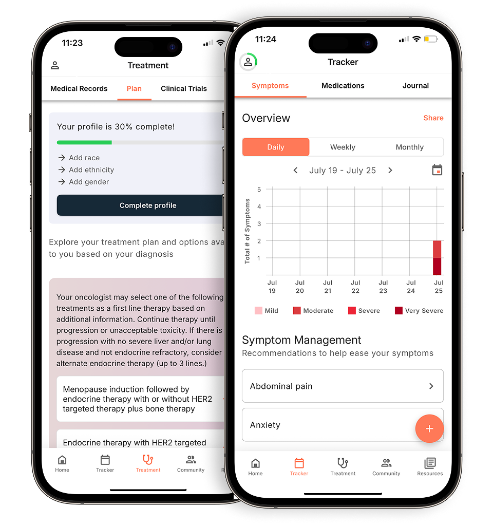
Cell differentiation plays a central role here. How well-differentiated a tumor’s cells are affects how actively they shed antigens like CA-125 into the bloodstream. Poorly differentiated, aggressive tumors tend to express more CA-125; well-differentiated mucinous or clear cell tumors often don’t.
A care team may rely heavily on CA-125 to monitor a high-grade serous case, yet find the marker nearly irrelevant when tracking a mucinous tumor. Understanding which subtype you have helps explain why your results look the way they do and sets the stage for examining how disease stage further shapes what this test can and can’t tell you.
Early stage vs. late stage detection
Stage is the single most reliable predictor of whether CA-125 will be elevated. Early-stage ovarian cancer is where this blood test most consistently falls short. CA-125 is elevated in only about 50% of Stage 1 cases, compared to roughly 90% of Stage 3 and 4 cases. When cancer is still confined to the ovary, relatively little antigen sheds into the bloodstream, meaning a normal result doesn’t rule anything out. That gap is precisely why catching and deciding how to treat ovarian cancer early remains so difficult. The tool most commonly associated with detection is least reliable at the moment it matters most. Understanding this limitation is part of why no single blood test tells the full diagnostic story, a point the next section addresses directly.
Moving beyond the blood test
A CA-125 result, whether elevated or within normal range, is never a definitive sign of ovarian cancer on its own. The actual diagnostic path combines a physical exam, imaging, and tissue analysis. Your care team will typically assess pelvic abnormalities first, then move to imaging before any surgical decision is made.
What many guides overlook is the necessity of a biopsy for definitive confirmation. Imaging can identify a suspicious mass, but only surgical pathology, examining the tissue directly, can confirm malignancy. CA-125 informs that process; it doesn’t replace it.
Two supplementary tools help your care team interpret ambiguous blood work more accurately. The HE4 test measures a second biomarker that’s often elevated when CA-125 is not, and the ROMA algorithm combines both CA-125 and HE4 markers with menopausal status to calculate a risk score. Together, they add important context, though they still don’t substitute for imaging. That’s where the next layer of assessment begins.
Imaging and physical assessment
When CA-125 results fall within a normal range, imaging and physical examination become the primary tools for identifying signs of ovarian cancer that blood work alone can’t reveal.
Transvaginal ultrasound (TVUS) is typically the first imaging step for assessing ovarian abnormalities. By positioning the ultrasound probe close to the ovaries, clinicians get a detailed look at adnexal masses, their size, texture, and whether they contain solid or fluid-filled components. These characteristics help distinguish benign cysts from suspicious growths that warrant further investigation.
When TVUS findings are inconclusive or a wider view is needed, CT scans or MRIs can detect potential metastasis in surrounding lymph nodes, the peritoneum, or distant organs. These modalities are particularly effective at mapping how far a tumor may have spread before surgery.
Pelvic examination rounds out this initial assessment. A clinician feeling for physical abnormalities, such as a fixed or irregular pelvic mass or the fluid accumulation known as ascites, can surface concerns that neither blood tests nor imaging fully capture on their own. Together, these three tools build a more complete clinical picture, setting the stage for the definitive step that imaging still can’t replace: surgical pathology.
The role of surgical pathology
Surgery remains the only definitive way to confirm an ovarian cancer diagnosis and determine its stage. No blood test or imaging result can replace it. Even when symptoms of ovarian cancer are present and a CA-125 result is elevated, a tissue sample reviewed by a pathologist is what transforms a clinical suspicion into a confirmed diagnosis. Pathologists may examine cell markers, including estrogen receptors, p53 mutations, and tumor grade, that CA-125 testing alone cannot provide information about.
Cancer care guidance for every step of your journey
Get treatment options, clinical trials, and support tailored to your diagnosis--all in one place.
Get started
See treatment options, manage symptoms, and stay informed—all in the app
View treatment options and trials personalized to your diagnosis—plus track progress in real time.
Continue in appDebulking, the surgical removal of as much tumor tissue as possible, also has a direct relationship with post-surgical CA-125 monitoring. After a successful debulking procedure, CA-125 levels typically fall, and your care team uses that trend to gauge how well treatment is working over time. Following CA-125 trends over time during and after treatment can help identify how well treatment is working, as an expected drop in CA-125 levels may not occur if disease remains present.
That said, it’s worth remembering that CA-125 can rise for reasons entirely unrelated to cancer, something the next section breaks down in detail.
Comparing CA-125 in cancer vs. benign conditions
A CA-125 result above 35 U/mL does not automatically point to ovarian cancer. The same protein elevation appears in a wide range of benign conditions, which is one of the most important reasons the test is never used alone.
The underlying mechanism explains why. When inflammation affects the peritoneum, the thin tissue lining the pelvic cavity, cells release CA-125 protein as part of a normal biological response. Ovarian cancer triggers that response, but so does endometriosis, pelvic inflammatory disease (PID), uterine fibroids, liver disease, and early pregnancy. Your care team will always weigh your result against your full clinical picture, not treat it as a standalone answer.
One of the most common questions providers hear is whether a high CA-125 signals ovarian cancer or endometriosis. Both can push levels well above 35 U/mL, and neither can be ruled in or ruled out by the number alone. What distinguishes malignancy is typically a rising trend over serial tests, a significantly elevated level (often above 200–300 U/mL in advanced disease), and imaging findings, factors that collectively support an ovarian cancer diagnosis and influence outcomes when detected early.
Understanding which condition is actually driving an elevated result requires the layered diagnostic approach covered in earlier sections, and the next section breaks down exactly what specific numerical ranges tend to look like across malignant versus benign causes.
Comparison: malignant vs. benign elevated CA-125
The pattern of CA-125 change matters far more than the absolute number in distinguishing ovarian cancer from other conditions.
A result of 54 or 66 U/mL sits above the standard 35 U/mL threshold, but that alone doesn’t point to cancer. Endometriosis is a noncancerous condition that can cause elevated CA-125 levels, while uterine fibroids are among the benign gynecologic conditions that can elevate CA-125 levels. Early-stage epithelial ovarian cancer may fall anywhere in a similar window, which is precisely why context matters. A number without a trend is just a number.
The differentiating factor is trajectory. A CA-125 that holds steady at 60 U/mL across three tests suggests a benign, stable source. The same level climbing from 40 to 60 to 85 U/mL over several months tells a different story, one your care team will take seriously as a potential signal of malignancy, even before imaging confirms anything.
That directional pattern, rising vs. stable, is what guides clinical decision-making far more reliably than any single reading. And it’s exactly why understanding your personal baseline matters as much as knowing the reference range, a point the next section explores in depth.
When a normal CA-125 is a critical baseline
A “normal” CA-125 result at diagnosis serves as a reference point that can shape every clinical decision that follows.
Before any therapy for ovarian cancer begins, that pretreatment number becomes your personal anchor. Even a reading of 12 U/mL, well within the standard 35 U/mL threshold, carries real weight once it’s documented. If that number climbs to 20 U/mL over subsequent tests, the absolute value still looks unremarkable on paper. But the direction tells a different story.
Trend over threshold. In practice, clinicians focus far less on where a number lands and far more on where it’s heading. A steady upward drift, even entirely within “normal” range, can prompt your care team to investigate before symptoms appear. This is the detail that rarely makes it into patient conversations but guides oncology decisions every day.
Monitoring treatment efficacy
A falling CA-125 after chemotherapy is one of the clearest signals your care team has that treatment is working, and a rising one can flag recurrence before you feel a thing.
When levels normalize following treatment, that’s a meaningful indicator of positive response. But the real clinical value comes from what happens next. Biochemical recurrence, a sustained rise in CA-125 confirmed across serial tests, typically precedes physical symptoms by months, giving your care team a critical window to act. Serial CA-125 testing during surveillance can help your doctor track patterns over time, which provides more information than a single test result. This gives you and your care team a critical early warning system, before symptoms become harder to manage.
Limitations and considerations for patients
A CA-125 result is a data point, not a verdict. Understanding its limits can protect you from unnecessary anxiety and misinterpretation.
Scanxiety is real. Waiting on marker results triggers significant emotional challenges for many patients, and a number that moves in either direction can feel all-consuming. That emotional weight is valid, but it’s also why context matters so much.
A few important caveats your care team will factor in:
- Different laboratories may use different reference ranges for CA-125 testing, so what is considered “normal” can vary depending on where your test is performed. To track trends accurately, use the same lab consistently throughout treatment.
- Early-stage and certain histologic subtypes, including mucinous and clear cell tumors, can present with completely normal markers even in advanced disease. This is why ovarian cancer and ultrasound imaging are typically used together rather than relying on CA-125 alone.
- CA-125 as a solo indicator isn’t clinically sufficient. Elevation or suppression only carries meaning when correlated with imaging, symptoms, and physical exam findings.
The test is a powerful tool within a personalized care plan, but it has edges. Recognizing those edges helps you ask sharper questions and interpret results with more clarity. That said, CA-125 isn’t the right tool for every clinical situation, and understanding when not to rely on it is just as important as knowing when it adds value.
When this test isn’t the right approach
CA-125 screening isn’t recommended for asymptomatic women at average risk, because using it outside appropriate clinical context can cause more harm than good.
The U.S. Preventive Services Task Force does not recommend routine CA-125 screening for asymptomatic women at average risk, and false positives are common with CA-125 screening in this population. A benign condition can elevate the marker, and without a corresponding abnormality, say, ovarian cancer on an ultrasound, that elevated result has no clear clinical meaning. Yet it often triggers invasive diagnostic follow-up, including surgeries that carry real complication risks for women who never had cancer.
For women with a strong family history or confirmed BRCA1/2 mutations, genetic testing, not CA-125, is typically the more appropriate starting point for assessing inherited risk. The right next steps to take will become clearer once you understand your specific risk profile, which is exactly what the key takeaways ahead are designed to help you frame.
Key takeaways: navigating your CA-125 results
A normal CA-125 result, below 35 U/mL, does not rule out ovarian cancer. What matters most is the full clinical picture: your symptoms, your family history, and what imaging on an ultrasound or CT scan reveals alongside your bloodwork. A single CA-125 test result alone is usually not very helpful. Your healthcare provider will order CA-125 tests in a series so they can identify changes in your levels over time. And a gynecologic oncologist is best positioned to interpret all of it together, within the context of your personalized care plan.
Disclaimer: The information provided in this article is for informational purposes only and is not intended to be a substitute for professional medical advice, diagnosis, or treatment. Outcomes4Me is not acting as your caregiver, and any suggestions or guidance offered should not replace the advice of your healthcare provider or qualified medical professional. Always seek the guidance of your physician or other qualified health provider with any questions you may have regarding a medical condition.
Personalized support for real care decisions
Understand your diagnosis, explore clinical trials, and track symptoms--all in one place.
Get started
Compare treatments, prepare for appointments, and track side effects—all in the app
Built for your diagnosis, Outcomes4Me gives you the tools to make confident, informed decisions—right when you need them.
Continue in app