
A diagnosis of ovarian cancer rarely arrives with immediate clarity. Surgery allows doctors to confirm your ovarian cancer stage through tissue analysis, which has been shown to be highly reliable for guiding treatment decisions.
Imaging tools like CT and MRI scans are valuable, but they have limitations in detecting small tumor deposits scattered across the abdominal lining. A scan might suggest a general picture, but it can’t tell your care team the full story. Surgery plays an important role in ovarian cancer staging, as tissue samples removed during surgery are used to determine your cancer’s pathological stage. This sets it apart from many other cancers where imaging alone determines the stage.
Understanding how that staging actually works is the natural next step, and that starts with the FIGO system.
Decoding FIGO staging. How far has it spread?
FIGO staging of ovarian cancer is the international standard for measuring how far the disease has spread, and it’s the framework your care team uses to build your personalized care plan. Established by the International Federation of Gynecology and Obstetrics, the FIGO system divides ovarian cancer into four stages, each reflecting a greater degree of spread from the original tumor site.
Stage I means the cancer is confined to one or both ovaries or fallopian tubes, with no spread beyond them. Stage II indicates the cancer has extended to nearby pelvic organs, the uterus, bladder, or rectum, but hasn’t traveled further. Stage III, which accounts for a significant proportion of diagnoses, involves spread to the abdominal lining (peritoneum) or to regional lymph nodes. And stage IV means the cancer has reached distant sites, most commonly the lungs, liver, or tissue outside the abdomen.
Staging helps your doctor determine the best treatment approach for you, which usually includes a combination of surgery and chemotherapy. A stage I diagnosis may require a very different treatment approach than a stage III, even if symptoms looked similar at the outset.
What makes this framework so consequential is that it can only be determined accurately through surgery. The surgical procedure removes visible tumors and provides the tissue samples needed for accurate staging. The surgical goal of removing as much disease as possible has its own clinical name and significance, which we’ll explore next.
Evidence-based guidance powered by NCCN Guidelines®
Personalized treatment plans shaped by the latest oncology standards—tailored to your diagnosis.
Get started
View your personalized treatment plan in the Outcomes4Me app
Use your diagnosis to unlock personalized NCCN Guidelines®-aligned recommendations.
Continue in app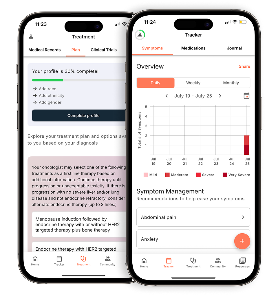
The goal of debulking: achieving optimal cytoreduction
Ovarian cancer debulking surgery has one clear objective. Remove as much tumor as possible to give every subsequent treatment the best possible foundation to work from.
Debulking, or cytoreduction, is the process of surgically removing tumor tissue throughout the abdomen and pelvis. Your care team aims for what’s called optimal debulking, leaving no visible tumor deposit larger than 1 centimeter. This threshold matters enormously. When residual disease falls below that benchmark, survival outcomes improve significantly, making this one of the most consequential goals in ovarian cancer treatment.
The reason comes down to biology. Chemotherapy works by targeting actively dividing cancer cells. Removing tumor burden through surgery may help reduce the number of cancer cells that need to be treated and could potentially improve how chemotherapy works in your body. Talk with your care team about how surgery fits into your overall treatment plan.
Even in stage IV, where cancer has spread beyond the abdomen to distant organs, surgery may still be on the table. For stage IV ovarian cancer, cytoreductive surgery may be considered as part of your treatment plan to help achieve optimal tumor removal. It’s a decision your care team will weigh carefully against your overall health and the extent of the disease.
What happens during the procedure: common surgical steps
Knowing what to expect after an ovarian cancer diagnosis starts with understanding exactly which procedures your care team may perform, and why each one matters for accurate staging.
Ovarian cancer staging surgery involves a coordinated set of steps designed to both treat and precisely map the disease. Here’s what that typically looks like:
Cancer care guidance for every step of your journey
Get treatment options, clinical trials, and support tailored to your diagnosis--all in one place.
Get started
See treatment options, manage symptoms, and stay informed—all in the app
View treatment options and trials personalized to your diagnosis—plus track progress in real time.
Continue in app- Total hysterectomy: A total hysterectomy typically involves removal of the uterus and cervix, though your surgical team can discuss exactly what will be removed in your procedure. This eliminates a primary site where cancer can establish or spread, and it’s a foundational step in most staging procedures.
- Bilateral salpingo-oophorectomy (BSO): Both ovaries and fallopian tubes are removed. Ovarian cancer can originate in the ovaries or fallopian tubes, and may spread between these structures or to other areas of the pelvis and beyond. Removing fallopian tubes alongside the ovaries is considered standard practice.
- Omentectomy: During ovarian cancer surgery, your doctor may remove the omentum, a tissue layer in the abdomen, as part of staging procedures to check for cancer spread. This is a routine and critical step in ovarian cancer surgery.
- Lymphadenectomy: Surgeons sample lymph nodes from the pelvis and abdomen. This procedure may help identify whether cancer has spread to the lymph nodes, which can be important for treatment planning. Lymph node sampling helps doctors determine whether ovarian cancer has spread, which is essential for accurate staging and treatment planning.
Recovery from this combination of procedures typically ranges from three to six weeks, depending on your overall health and the extent of surgery involved. Each of these steps directly informs what comes next, including the pathology report, which will confirm your final stage and grading.
The pathology report. Your final stage and grading
Your ovarian cancer stage isn’t confirmed until a pathologist has analyzed the tissue removed during surgery. What you receive after surgery is a pathology report, and it contains two separate but equally important findings. Staging describes how far the cancer has spread. The difference between ovarian cancer stage I vs. stage IV outcomes often depends on what this report reveals about tumor distribution beyond the ovaries. Grading is one way doctors assess how the cancer cells appear under a microscope, which can help inform treatment planning. Lower-grade cells closely resemble healthy cells and tend to grow more slowly, while higher-grade cells look more abnormal and behave more aggressively.
Both pieces of information directly shape what happens next. Your care team will use your confirmed stage and grade together to determine whether adjuvant chemotherapy, treatment given after surgery to eliminate remaining cancer cells, is appropriate for your personalized care plan. A stage I, low-grade tumor may require little to no additional treatment, while many women with higher-stage ovarian cancer receive systemic therapy (chemotherapy) after surgery, and chemotherapy is usually recommended for higher-grade tumors.
And because these findings carry so much weight, keeping a digital copy of your pathology report is a practical step worth taking. It ensures you’re equipped for second opinions, enables clearer conversations with specialists, and gives you accurate information if you explore clinical trials down the line.
What you need to know
Surgery is the cornerstone of both treating and staging ovarian cancer, and the decisions made in the operating room directly shape every treatment step that follows.
Accurate staging can’t happen without surgery. Procedures like omentectomy and lymph node dissection serve both therapeutic and diagnostic purposes. The tissue your care team removes is what allows a pathologist to confirm your final FIGO stage, and that confirmed stage unlocks access to the most appropriate, personalized care plan for your specific situation.
Here’s what’s worth keeping in mind as you process everything covered in this article:
- Specialist matters. Patients treated by a specialized gynecologic oncologist have significantly better outcomes, making your choice of surgeon one of the most consequential decisions you’ll face.
- Optimal debulking is the goal. Leaving less than 1 cm of residual tumor is the benchmark your care team aims for, and it’s directly tied to prognosis.
- Your FIGO stage is confirmed post-surgery. Imaging gives an estimate; the pathology report gives the answer.
- Staging data drives what comes next. Confirmed stage determines which therapies your doctor may recommend, since ovarian cancers at different stages are treated differently.
Your surgical and staging data forms the foundation of every treatment decision ahead of you. The more precisely your stage is defined, the better positioned you are to access treatments and clinical trials matched to your exact diagnosis.
Download the Outcomes4Me app to access personalized, evidence-based resources that put you in control of navigating your condition — because better outcomes start with better information.
Disclaimer: The information provided in this article is for informational purposes only and is not intended to be a substitute for professional medical advice, diagnosis, or treatment. Outcomes4Me is not acting as your caregiver, and any suggestions or guidance offered should not replace the advice of your healthcare provider or qualified medical professional. Always seek the guidance of your physician or other qualified health provider with any questions you may have regarding a medical condition.
Personalized support for real care decisions
Understand your diagnosis, explore clinical trials, and track symptoms--all in one place.
Get started
Compare treatments, prepare for appointments, and track side effects—all in the app
Built for your diagnosis, Outcomes4Me gives you the tools to make confident, informed decisions—right when you need them.
Continue in app